Hormesis is a two-phased dose-response relationship whereby low-dose exposures have a beneficial effect and high-doses toxic. Pulsed ElectroMagnetic (very low) Frequency (PEMF) mitohormesis is the slight generation of reactive oxygen species that activates the transient receptor potential C1 (TRPC1) Ca2+ channel.
Franco-Obregón A, Tai YK, Wu KY, Iversen JN, Wong CJK. The Developmental Implications of Muscle-Targeted Magnetic Mitohormesis: A Human Health and Longevity Perspective. Bioengineering (Basel). 2023 Aug 12;10(8):956. PMC free paper
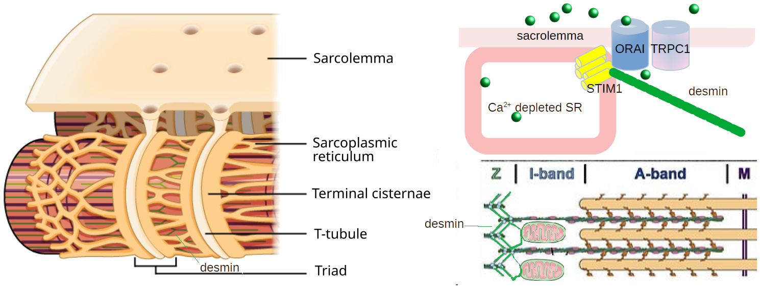
Graphical Summary ,,,,,Ca2+ signaling and mitochondrial adaptations are shared between magnetic fields and endurance training in the development of oxidative muscles. Ca2+ feedback from TRPC1 to the mitochondria is part of this process; Ca2+ modulates mitochondrial respiration. This post makes a case for the intermediate filament protein desmin as a physical connection.
The origin of this investigation
In 2022 a QuantumTx cultured myocyte study demonstrated that the mitohormesis effect was greater when the direction of the magnetic field was in the same direction of gravity. See the gravity and PEMF post. Why? Plants have intracellular structures called statoliths that respond to gravity and tug on actin filaments in the plant cytosol. These same actin filaments are connected to membrane rafts that contain Ca2+ cannels.
Wong CJK, Tai YK, Yap JLY, Fong CHH, Loo LSW, Kukumberg M, Fröhlich J, Zhang S, Li JZ, Wang JW, Rufaihah AJ, Franco-Obregón A. Brief exposure to directionally-specific pulsed electromagnetic fields stimulates extracellular vesicle release and is antagonized by streptomycin: A potential regenerative medicine and food industry paradigm. Biomaterials. 2022 Aug;287:121658. PMC free paper
Wong 2022 laid the foundation of the healing extracellular vesicle release in those seniors benefitting from BIXEPS sessions. There appears to be something autocrine or paracrine going on. Wong 2022 looked at C2C12 cultured myoblasts and myotubules. These cells experience gravity just like the roots of plants. One would think that this is nothing like the force experience by fully differentiated myocytes in contracting muscle fibers. Could gravity and everyday forces be influencing the way thin, thick, and intermediate filaments are organized?
TRPC1 and eccentric muscle contraction
Eccentric contractions are generated by an external load on the muscle; the muscle fibers lengthen as they contract. Eccentric contraction of the bicep involves load bearing and fiber lengthening as the weight is lowered. Putting the brake on knee movement while barreling down the stairs is an example of eccentric contraction of the quadricep.
Zhang BT, Whitehead NP, Gervasio OL, Reardon TF, Vale M, Fatkin D, Dietrich A, Yeung EW, Allen DG. Pathways of Ca²⁺ entry and cytoskeletal damage following eccentric contractions in mouse skeletal muscle. J Appl Physiol (1985). 2012 Jun;112(12):2077-86. Free paper A quote from the abstract:
“Muscles that are stretched during contraction (eccentric contractions) show deficits in force production and a variety of structural changes, including loss of antibody staining of cytoskeletal proteins. Extracellular Ca2+ entry and activation of calpains have been proposed as mechanisms involved in these changes. The present study used isolated mouse extensor digitorum longus (EDL) muscles subjected to 10 eccentric contractions and monitored force production, immunostaining of cytoskeletal proteins, and resting stiffness. Possible pathways for Ca2+ entry were tested with streptomycin (200 μM), a blocker of stretch-activated channels, and with muscles from mice deficient in the transient receptor potential canonical 1 gene (TRPC1 KO), a candidate gene for stretch-activated channels. At 30 min after the eccentric contractions, the isometric force was decreased to 75 ± 3% of initial control and this force loss was reduced by streptomycin but not in the TRPC1 KO. Desmin, titin, and dystrophin all showed patchy loss of immunostaining 30 min after the eccentric contractions, which was substantially reduced by streptomycin and in the TRPC1 KO muscles. Muscles showed a reduction of resting stiffness following eccentric contractions, and this reduction was eliminated by streptomycin and absent in the TRPC1 KO muscles. Calpain activation was determined by the appearance of a lower molecular weight autolysis product and μ-calpain was activated at 30 min, whereas the muscle-specific calpain-3 was not. To test whether the loss of stiffness was caused by titin cleavage, protein gels were used but no significant titin cleavage was detected. These results suggest that Ca2+ entry following eccentric contractions is through a stretch-activated channel that is blocked by streptomycin and encoded or modulated by TRPC1.”
In this post evidence is presented that desmin is not only part of the Z-line, the part of the sacromer where actin thin filaments attach, but is also a binder of mitochondria and the sensor of Ca2+ depletion from the sarcoplasmic reticulum STIM1. Was desmin and titin degradation a feedback mechanism to prevent muscle over use?
The complicated interplay between STIM1 and TRPC1
This section of the post will build the case for the potential of desmin to modulate TRPC1 indirectly through STIM1.
TRPC1 interacts with STIM1
Ambudkar IS, de Souza LB, Ong HL. TRPC1, Orai1, and STIM1 in SOCE: Friends in tight spaces. Cell Calcium. 2017 May;63:33-39. PMC free paper A direct quote from the abstract, arranged into bullet points:
“Store-operated calcium entry (SOCE) is a ubiquitous Ca2+ entry pathway that is activated in response to depletion of ER-Ca2+ stores and critically controls the regulation of physiological functions in miscellaneous cell types.
- The transient receptor potential canonical 1 (TRPC1) is the first member of the TRPC channel subfamily to be identified as a molecular component of SOCE.
- While TRPC1 has been shown to contribute to SOCE and regulate various functions in many cells, none of the reported TRPC1-mediated currents resembled ICRAC, the highly Ca2+-selective store-dependent current first identified in lymphocytes and mast cells.
- Almost a decade after the cloning of TRPC1 two proteins were identified as the primary components of the CRAC channel. The first, STIM1, is an ER-Ca2+ sensor protein involved in activating SOCE. The second, Orai1 is the pore-forming component of the CRAC channel.
- Co-expression of STIM1 and Orai1 generated robust ICRAC.
- Importantly, STIM1 was shown to also activate TRPC1 via its C-terminal polybasic domain, which is distinct from its Orai1-activating domain, SOAR.
- In addition, TRPC1 function critically depends on Orai1-mediated Ca2+ entry which triggers recruitment of TRPC1 into the plasma membrane where it is then activated by STIM1.
- More importantly, TRPC1 and Orai1 form discrete STIM1-gated channels that generate distinct Ca2+ signals and regulate specific cellular functions.
- Surface expression of TRPC1 can be modulated by trafficking of the channel to and from the plasma membrane, resulting in changes to the phenotype of TRPC1-mediated current and [Ca2+]i signals. Thus, TRPC1 is activated downstream of Orai1 and modifies the initial [Ca2+]i signal generated by Orai1 following store depletion.
This review will summarize the important findings that underlie the current concepts for activation and regulation of TRPC1, as well as its impact on cell function.”
Conte, E.; Imbrici, P.; Mantuano, P.; Coppola, M.A.; Camerino, G.M.; De Luca, A.; Liantonio, A. Alteration of STIM1/Orai1-Mediated SOCE in Skeletal Muscle: Impact in Genetic Muscle Diseases and Beyond. Cells 2021, 10, 2722. free paper
For those that like good diagrams…
- Fig 1 The stromal interacting molecule (STIM1) spans the sacroplasmic reticulum, where Ca2+ is stored, and the plasma membrane, where Ca2+ channels can be recruited. The EF hand is a Ca2+ binding motif. Desmin interacts with STIM1’s CC1 domain, see the next section. The K-rich stuff is claimed to activate TRPC1 by Ambudkar 2017.
- Fig 2 Ca2+ release-activated Ca2+ channel (CRAC channel) and are considered the major Store-operated Ca2+ entry SOCE in skeletal muscle cells. Orai is a plasma membrane Ca2+ channel that interacts with STIM1.
- Fig 3 is a diagram of TRPC1/Orai/STIM1. Note the red/yellow STIM1 molecule and its interaction with TRPC1. Imagine a desmin filament binding to this red domain and acting as a conduit for Ca2+ entering throug TRPC1… and ultimately directing Ca2+ to the mitochondria.
- Fig 4 a flow charts of the proposed sequence of events and ways of modulating STIM. Desmin was not included in this figure.
Desmin interacts with STIM1
Zhang H, Bryson VG, Wang C, Li T, Kerr JP, Wilson R, Muoio DM, Bloch RJ, Ward C, Rosenberg PB. Desmin interacts with STIM1 and coordinates Ca2+ signaling in skeletal muscle. JCI Insight. 2021 Sep 8;6(17):e143472. Free paper
“Stromal interaction molecule 1 (STIM1), the sarcoplasmic reticulum (SR) transmembrane protein, activates store-operated Ca2+ entry (SOCE) in skeletal muscle and, thereby, coordinates Ca2+ homeostasis, Ca2+-dependent gene expression, and contractility. STIM1 occupies space in the junctional SR membrane of the triads and the longitudinal SR at the Z-line.
How STIM1 is organized and is retained in these specific subdomains of the SR is unclear.
Here, we identified desmin, the major type III intermediate filament protein in muscle, as a binding partner for STIM1 based on a yeast 2-hybrid screen. Validation of the desmin-STIM1 interaction by immunoprecipitation and immunolocalization confirmed that the CC1-SOAR domains of STIM1 interact with desmin to enhance STIM1 oligomerization yet limit SOCE.
Based on our studies of desmin-KO mice, we developed a model wherein desmin connected STIM1 at the Z-line in order to regulate the efficiency of Ca2+ refilling of the SR. Taken together, these studies showed that desmin-STIM1 assembles a cytoskeletal-SR connection that is important for Ca2+ signaling in skeletal muscle. “
This study tested the hypothesis that desmin associates with the carboxy-terminus of STIM1-SOAR (STIM1-CT, 238–535 aa) in order to influence STIM1 oligomerization, a mechanism important for STIM1’s actions on Orai1.
The same domain in desmin was found not to interact with TRPC1.
Desmin and the mitochondria
Abstracts from two representative publications demonstrating interaction f desmin filaments with mitochondria are quoted:
Milner 2000
Milner DJ, Mavroidis M, Weisleder N, Capetanaki Y. Desmin cytoskeleton linked to muscle mitochondrial distribution and respiratory function. J Cell Biol. 2000 Sep 18;150(6):1283-98. free paper
“Ultrastructural studies have previously suggested potential association of intermediate filaments (IFs) with mitochondria. Thus, we have investigated mitochondrial distribution and function in muscle lacking the IF protein desmin.
- Immunostaining of skeletal muscle tissue sections, as well as histochemical staining for the mitochondrial marker enzymes cytochrome C oxidase and succinate dehydrogenase, demonstrate abnormal accumulation of subsarcolemmal clumps of mitochondria in predominantly slow twitch skeletal muscle of desmin-null mice.
- Ultrastructural observation of desmin-null cardiac muscle demonstrates in addition to clumping, extensive mitochondrial proliferation in a significant fraction of the myocytes, particularly after work overload.
- These alterations are frequently associated with swelling and degeneration of the mitochondrial matrix. Mitochondrial abnormalities can be detected very early, before other structural defects become obvious.
- To investigate related changes in mitochondrial function, we have analyzed ADP-stimulated respiration of isolated muscle mitochondria, and ADP-stimulated mitochondrial respiration in situ using saponin skinned muscle fibers. The in vitro maximal rates of respiration in isolated cardiac mitochondria from desmin-null and wild-type mice were similar.
- However, mitochondrial respiration in situ is significantly altered in desmin-null muscle. Both the maximal rate of ADP-stimulated oxygen consumption and the dissociation constant (K m) for ADP are significantly reduced in desmin-null cardiac and soleus muscle compared with controls.
- Respiratory parameters for desmin-null fast twitch gastrocnemius muscle were unaffected. Additionally, respiratory measurements in the presence of creatine indicate that coupling of creatine kinase and the adenine translocator is lost in desmin-null soleus muscle. This coupling is unaffected in cardiac muscle from desmin-null animals. All of these studies indicate that desmin IFs play a significant role in mitochondrial positioning and respiratory function in cardiac and skeletal muscle.
it is suggested that the absence of desmin leads to loss of proper mitochondrial positioning which might be the cause of the observed compromised mitochondrial function. As a consequence, cell death follows either directly due to energy deprivation or/and indirectly due to oxidative stress caused by mitochondrial proliferation and eventually degeneration and direct release of death triggering factors.”
Smolina 2014
Smolina N, Bruton J, Sjoberg G, Kostareva A, Sejersen T. Aggregate-prone desmin mutations impair mitochondrial calcium uptake in primary myotubes. Cell Calcium. 2014 Oct;56(4):269-75. PubMed
“Desmin, being a major intermediate filament of mature muscle cell, interacts with mitochondria within the cell and participates in mitochondria proper localization. The goal of the present study was to assess the effect of aggregate-prone and non-aggregate-prone desmin mutations on mitochondrial calcium uptake.
- Primary murine satellite cells were transduced with lentiviruses carrying desmin in wild type or mutant form, and were induced to differentiate into myotubes.
- Four mutations resulting in different degree of desmin aggregates formation were analyzed.
- Tail domain mutation Asp399Tyr has the mildest impact on desmin filament polymerization, rod domain mutation Ala357Pro causes formation of large aggregates composed of filamentous material, and Leu345Pro and Leu370Pro are considered to be the most severest in their impact on desmin polymerization and structure.
- For mitochondrial calcium measurement cells were loaded with rhod 2-AM.
We found that aggregate-prone mutations significantly decreased Ca2+mit, whereas non-aggregate-prone mutations did not decrease Ca2+mit. Moreover aggregate-prone desmin mutations resulted in increased resting cytosolic [Ca(2+)]. However this increase was not accompanied by any alterations in sarcoplasmic reticulum calcium release. We suggest that the observed decline in Ca2+mit was due to desmin aggregate accumulation resulting in the loss of desmin mitochondria interactions.”
Desmin as a conduit for Ca2+ ?
An attempt was made to find crystal structures of polymerized desmin just to determine the surface charge and whether they have a net negative surface charge that would support a double layer of cations. None were found. While this post is not addressing deminopathies, the images in the Clemen 2013 review offers some excellent insights into how demin might be contributing to mitohormesis.
Clemen CS, Herrmann H, Strelkov SV, Schröder R. Desminopathies: pathology and mechanisms. Acta Neuropathol. 2013 Jan;125(1):47-75. PMC free paper.
- Fig 1 This is a difficult but exciting figure showing H&E staining (Panel A), aggregated desmin (Panel B), and a spot of the myofibril that does not have cytochrome C oxidase activity (panel C). The difficulty is for those not used to looking at stainings of enzmatic activity.
- Fig 2 Indirect immunofluorescence labeling of desmin and αB-crystallin, a chaperone protein that refolds misfolded proteins, in a desminopathy. Note the presence of sarcoplasmic and subsarcolemmal pathological protein aggregates.
- Fig 3 is a really nice set of scanning electron micrographs showing misfolded desmin between the extracellular matrix and the sarcomers. The Z-lines are visible. The telling things about these images is that we know that TRPC1 is somewhere between the misfolded desmin and the extracellular matrix. Things are pretty crowded.
- Fig 4 and table 1 describe the location of mutations in desmin that affect muscle function. If there is TERPC1 to mitochondria communication via desmin, do these mutations affect it?
- Fig 5 describes the making of a desmin filament from monomers to tetramers to filaments….
- Fig 6 illustrates both organized and desminopathy Z-disks. Damaged and good mitochondria are shown surronding the Z-disks along with desmin fibrils. Myonuclei are mentioned in the text but unfortunately, not shown in this fantastic cartoon. What does this mean for CRY that also makes its way to the nuclei as a transcription factor?
- Fig 7 is a a dual fluorescent immunohistochemistry of desmin and the less well known intermediate filament syncoilin. The Z-lines and clumps of intermediate filaments are pretty visible in this image of a desminopathy.
- Fig 8 are some images of mutant desmins expressed in 3T3 fibroblasts and colocalization of vimentin, an intermediate filament naturally found in fibroblasts. Nuclei are stained blue with DAPI, very dramatic desmin and nuclei orientation.
- Fig 9, mutant desmins do not polymerize correctly in vitro.
Nothing in this review answers the question of desmin fibrils being a conduit for Ca2+ in the PEMF stimulated myocyte. Chlemin 2013 might raise the question, “Could PEMF do anything to treat desminopathies?”
Light is needed for the downfield response in C2C12 cells
Iversen JN, Tai YK, Wu KY, Wong CJK, Lim HY, Franco-Obregón A. Magnetically Stimulated Myogenesis Recruits a CRY2-TRPC1 Photosensitive Signaling Axis. Cells. 2025 Feb 6;14(3):231. free paper
Iversen 2025 made the case for cryptochrome 2 being a source of reactive oxygen species in the presence of light and magnetic fields. The downfield is better than upfield PEMF disappeared in cultured C2C12 cells in the dark. This work also demonstrated direct binding of Cry2 to TRPC1. The premise is that direct binding means that ROS do not have to diffuse far to react with TRPC1 or surrounding lipids in such a way as to influence channel gating. A post CRY2/TRPC1 was devoted to this publication last year. This model requires the presence of “biophotons” that was the subject of a post last year.
- Could gravity influence the direction of desmin intermediate filaments in C2C12 cultured cells?
- Could muscle fiber stretching and influence the layout of desmin?
- Could PEMF do more in terms of mitorhormesis than just generating ROS from the mitochondria and/or CRY2?
- If there is a Ca2+ counterion cloud on desmin, could a moving magnetic field set these charge in motion too via Faraday’s Law of induction?
Leave a Reply