This post examines the effect of up field and down field static magnetic fields in plants and pulsed electromagnetic fields in a a skeletal muscle cell line. Up field and down field is relative to the force of gravity in a manner distinct from the magnetic field of the earth. Just how do these two seemingly distinct physical forces combine in plants and animals?
Related posts
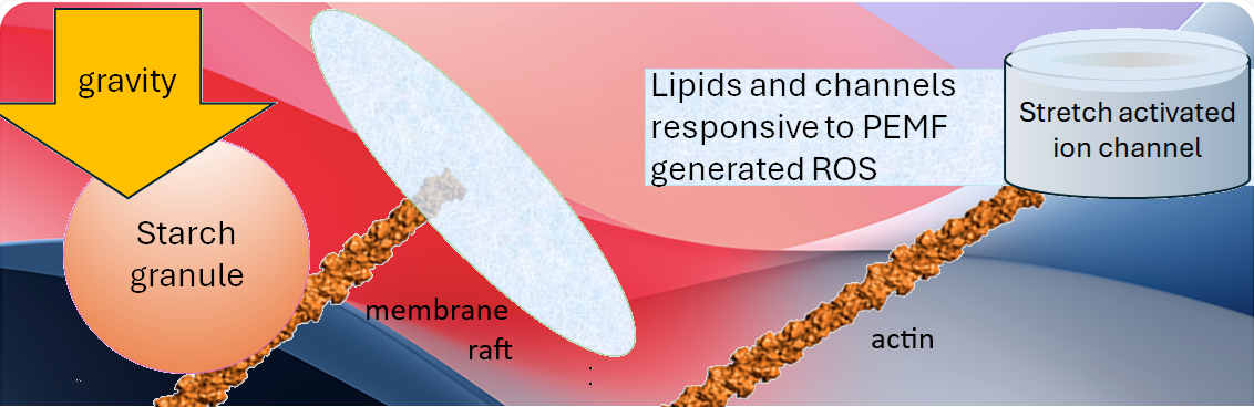
Water and the putative gravity sensor starch, are diamagnetic. This post will stick to the featured image and find support for this model of gravity/PEMF synergy.
- , exclusion zone water looks at the ordering of water in magnetic fields owing the the hydronium H3O+ cation.
- Magnetic fields and water looks at the influence of static magnetic fields and and the interaction of ions and water.
- Triplet oxygen examines the interaction of magnetic fields in with unpaired electrons in triplet oxygen.
- Stretch activated TRPC1 is an ion channel even found in adipocytes.
- PEMF and Sphingosine modulate TRPC1 in skeletal muscle. This is a lipid raft lipid.
How plants sense gravity
Just to not forget basic physics, Archimedes Principle states that any object in water in a confined space will “try” to displace water exerting a force somewhere in the cell.

The Wikipedia authors have built an interesting collection of anecdotal examples of Archimedes Principle and buoyant forces. A helium balloon in a car turning or accelerating… Lorentzian forces from PEMF on a statolith/amyloplast with negative surface charges…

This image explains statoliths/amyloplasts interacting with actin contecting to plant cell membrane rafts, connected to stretch activated ion channels that might have some homology to TRPC1. These spheres could conceivably be any dense organelle connecting to the cell membrane by actin or another filament.
falling amyloplasts/statoliths
Gravitropism is the term used by wikipedia authors to describe the role of the plant hormone auxin in gravity sensitive amyloplasts that press against actin connected to mechano sensitive ion channels. Biology Insights claims that these structures are dense and contain enzymes.
stretch activated ion channels
Perrin RM, Young LS, Murthy U M N, Harrison BR, Wang Y, Will JL, Masson PH. Gravity signal transduction in primary roots. Ann Bot. 2005 Oct;96(5):737-43. . PMC free paper
Perrin 2005 claimed no plant homologs of mammalian mechanosensitive TRP channels, but some domain homology to bacterial mechanosensitive channels in bacteria. Auxin transporting PIN proteins were also mentioned.
lipid rafts
Kordyum EL, Artemenko OA, Hasenstein KH. Lipid Rafts and Plant Gravisensitivity. Life (Basel). 2022 Nov 7;12(11):1809. ; PMC free paper
The actin cytoskeleton, attachment to cholesterol rich membrane raft domains and Ca2+ were mentioned. Clinostat data were cited in this review. A clinostat is a rotating device that negates the effect of gravity on root tropism.
| fat | static | clinorotated |
|---|---|---|
| saturated | 43.5% | 64.5% |
| monoenoic | 6.5% | |
| polyenoic | 29%, | |
| polyenoic fatty acids | 28.2% | 28.2% |
| cholesterol | 1x | 7x |
, and the content of saturated fatty acids increased (66.7%), monoenoic fatty acids decreased. Literature cited in this paper reported higher proportions of sterols, sphingolipids, and saturated phospholipids were also detected in the microdomains of oats and rye in comparison with those in the cell membrane.
Activation of kinases
Chen J, Yu R, Li N, Deng Z, Zhang X, Zhao Y, Qu C, Yuan Y, Pan Z, Zhou Y, Li K, Wang J, Chen Z, Wang X, Wang X, He SN, Dong J, Deng XW, Chen H. Amyloplast sedimentation repolarizes LAZYs to achieve gravity sensing in plants. Cell. 2023 Oct 26;186(22):4788-4802.e15. . PMC free paper
- Gravity pushes amyloplasts against plasma membrane covered with LAZY proteins. LAZY proteins are localized to lipid raft domains.
- , MKK5–MPK3 MAPK protein kinase family members phosphorylates LAZY proteins. MKK5 is upstream of MPK3. UniProt indicates both are modulated by hydrogen peroxide. LAZY4 shows stronger interaction with MPK3 and MKK5 after gravistimulation in Arabidopsis.
- Phosphorylation promotes LAZY proteins to enrich on the surface of amyloplasts via interacting with the Transport of Chloroplast proteins.
- Polar auxin transport (PAT) that asymmetrically distributes auxin. ATP fueled P-Glyco Proteins (PGP) and PIN proteins that transport the anionic form of auxins.
- Secreted auxin, of which indol-3-acetic acid is the most abundant, acts as a growth hormone.
starches settle with gravity
These are some Wikimedia Commons images of hydrated tapioca starch balls settling with gravity in a popular drink.

That these starch particles settle suggest they are more dense than water.
Kuznesov & Hasenstein1996: starch is diamagetic?
Kuznetsov OA, Hasenstein KH. Intracellular magnetophoresis of amyloplasts and induction of root curvature. Planta. 1996;198(1):87-94. Sci-hub free paper

A combination of Kuzetsov & Hohenstein 1996 Fig 1A showing magnetic field lines around a wedge shaped permanent magnet and repulsion of 10 μm starch granule in water in a 0.5 mm microcapillary tube. Oersted (Oe) units were used to describe the force of the magnetic field. Fig 3 reports 4 kOe. “he H-field strength inside a long solenoid wound with 79.58 turns per meter of a wire carrying 1A is approximately 1 Oersted. ” The 78.58 turns comes from 100/4π.
This post is not going to get into some very complicated images of amyloplast redistribution and response there of to mechanical forces in flax seedlings.
A plant static magnetic field experiment
Jin Y, Guo W, Hu X, Liu M, Xu X, Hu F, Lan Y, Lv C, Fang Y, Liu M, Shi T, Ma S, Fang Z, Huang J. Static magnetic field regulates Arabidopsis root growth via auxin signaling. Sci Rep. 2019 Oct 7;9(1):14384. PMC free paper
In these studies the N-S pole of the static magnet was orientated 0o down towards the center of the earth or 180o up. These authors “grew Arabidopsis seedlings in petri dishes that were closely attached to the side of a magnet, which is a cube with each side length of 10 cm and produces around 600 mT magnetic field at the surface of the north (N) and south (S) poles. Considering that petri dishes contained the solid medium and the roots actually grew about 5 mm away from the magnet surface, we measured magnetic fields 5 mm away from the magnet surface. Magnetic intensities at the side in parallel with the direction of magnetization ranged from 580 mT to 420 mT with the highest close to the poles and the lowest at the middle between the two poles, while at the side of the N or S pole ranged from 550 mT to 460 mT with the highest at the edge and the lowest at the center.”
North pole at 0o and 90o but not North 180o promotes increases in root length. These are just some top responding snap shots.
| gene ontology change | roots 0o North | roots 180o North |
| increase | p~0.01, >20 oxidation reduction genes | p~0.0001 response to growth regulators karrikin, 9 genes |
| decrease | p~0.01, >20 oxidation reduction genes | mention YUK9 a gene of auxin synthesis |
North 0o -promoted root growth is regulated by auxin signaling.
- Death receptor 5 (DR5) is a transcription factor that promotes transcription of genes related to programmed cell death apoptosis. A promoter for this TF was fused to Green Fluorescent Protein GFP). North 0o (down field) decreased green fluorescence.
- The auxin efflux carrier 3 promoter of PIN3 was also fused to the GFP gene. N0o down field almost halved the fluorescent signal implying that the ability to transport auxin out of the root cells was also increased.
- The promoter of the AUX1 transporter was fused to a Yellow Fluorescence Protein (YFP) gene. N0oalmost 2x yellow fluorescence.
- “The cry1 cry2 double mutant produced significant shorter roots than WT under the local geomatical field (CK), indicating that CRYs are positive regulators for primary root growth. Root length were very similar between cry1 cry2 seedlings treated with and without N0, while that of WT was significantly longer in N0 than in CK. These results suggest that CRYs play a role in root responses to SMF.”
In mammalian muscle cells
We do not have 100 years of research on how mammalian muscle cells sense gravity and if there is an interaction with magnetic fields, static or pulsed. The interesting thing to note is that mammalian muscle cells have glycogen granules.

The glycogen diamagnetic rabbit hole will be avoided. The featured image is to focus on the concept that glycogen granules and organelles that settle relative to the cytosol might be activating stretch activated channels that are activated by different means by reactive oxygen species generated by PEMF.
Starch/glycogen granules mammalian muscle cells
Prats C, Graham TE, Shearer J. The dynamic life of the glycogen granule. J Biol Chem. 2018 May 11;293(19):7089-7098. PMC free paper
- 10-40 nm diameter
- 1-2% tissue weight
- actin rich structures at I-band of skeletal muscle. The I-band is where the actin portion of the actomyosin complex attaches.
This post will not go into great detail of the Prat 2018 review. Suffice it to say, there is some overlap between glycogen granules in mammalian skeletal muscle and starch granules in root tips. No evidence was found on the response of glycogen granules to gravity. Some literature on PubMed was found on use of glycogen radio frequency magnetic resonance imaging. (MRI)
Ríos E, Samsó M, Figueroa LC, Manno C, Tammineni ER, Rios Giordano L, Riazi S. Artificial intelligence approaches to the volumetric quantification of glycogen granules in EM images of human tissue. J Gen Physiol. 2024 Sep 2;156(9):e202413595. PMC free paper
- Glycogen granules in skeletal muscle are a factor of diet, physiology, and pathophysiology.
- AI models were developed to derive granular glycogen content from electron-microscopic images of human muscle.
- The intermyofibrillar spaces and the sarcomeric I band had the highest granular content. The measured glycogen concentration was low enough to allow for a substantial presence of non-granular glycogen.
- The goal was to develop 3D approximations from 2D images.
- The volume content was around 3%

Prats has a transmission electron microscope image that places a large granule Rios had some examples in which starch granules were around the sarcoplasmic reticulum.
glycogen granules in skeletal muscle
Ørtenblad N, Nielsen J. Muscle glycogen and cell function–Location, location, location. Scand J Med Sci Sports. 2015 Dec;25 Suppl 4:34-40. Sci-Hub free paper
A few interesting comments from this review.
- The high concentration of macromolecules give it a high density and a gel like structure with poor diffusion. This is interesting. Is this stuff more or less dense than glycogen granules meaning do glycogen granules boant and respond o gravity with an upward force?
- Subsarcolemmal glycogen is located from the outermost myofibril to the
- sarcolemma. Is this close to TRPC1?
- intermyofibrillar glycogen is located between myofibrils. This is the predominant location in human muscle.
- Intramyofibrillar glycogen is within the myofibrils
- A connection between intramyofibrillar glycogen and SR Ca2+release is related to muscle fatigue. Intramyofibrillar glycogen is depleted first during prolonged exercise.
- Mitochondria, sources of ATP and reactive oxygen species, are beneath the sarcolemma and between the myofibrils, mainly near the I-band, the same location of the glycogen granules based on other reports.

On a very speculative level, one would expect viscosity, density, and the structure of water to differ. Hydrolysis of glycogen would only add to the variability.
In cultured C2C12
Wong CJK, Tai YK, Yap JLY, Fong CHH, Loo LSW, Kukumberg M, Fröhlich J, Zhang S, Li JZ, Wang JW, Rufaihah AJ, Franco-Obregón A. Brief exposure to directionally-specific pulsed electromagnetic fields stimulates extracellular vesicle release and is antagonized by streptomycin: A potential regenerative medicine and food industry paradigm. Biomaterials. 2022 Aug;287:121658. free paper
Instead of the concept of a static magnetic field (downward) we have the concept of a 15 or 50Hz pulsed magnetic field of 1.5 mT on the secretion of myokines. Note that this is over 100x less field intensity. Here “down” means 0o relative to the force of gravity. “Up” is the same as “180o” in the plant study. Instead of changing the orientation of the permanent magnet, the direction of the current flow of the Helmholtz coil was changed.
- Fig 1 examined the influence of direct downfield 1.5 mT PEMF exposure of myoblasts on the surface culture dishes or in suspension. Cell numbers The general theme is that like plants and secreted auxin, the PEMF effect is manifested by secretion of “myokines” into the medium.
- Fig S1 examined the effect of turning the culture flasks on the sides such that gravity down pushed the contents of the cells to one end. PEMF down was defined as towards the bottom of the culture flask such that the current flow was along long axes of the cells.
- Fig 2 Like static magnetic fields in plants, downward directed PEMF is more effective in increasing the cell number than upward (180o) and horizontal PEMF.
- Fig 3 No parallels in the flax seedling. Tested the ability of common mammalian cell culture antibiotic, streptomycin to block the effect of PEMF. It is known that aminoglycoside antibiotics block the stretch activated TRPC1 channels.
- Fig 4, direct down field PEMF exposure increased transcripts of a list of TRP channels, Cyclins B1 and D1. relative to up field PEMF and no PEMF exposure.
- Fig 5, Down field PEMF conditioned medium increased transcripts of a list of TRP channels, Cyclins B1 and D1. relative to up field PEMF and no PEMF.
- Fig 6 Reactive oxygen species generation was measured in C2C12 myoblasts exposed to up and down PEMF. Down generated more ROS than up even though the current flow would be along the attachment surface. Down PEMF conditioned medium promoted increases in cell number but not ROS generation.
- Fig 7 The administration of downward magnetically-conditioned media harvested from differentiated myotubes, promoted the differentiation of proliferating myoblasts and forestalled proliferation, Indeed up PEMF increased cyclins B1 and D1 relative to no exposure and down PEMF. Down PEMF increased relative amounts of phosphorylated ERK and decreased relative amounts of phosphorylated JNK kinases, ERK and JNK are members of the MAPK superfamily of kinases bringing us back to pathways of plant gravity sensing and auxin secretion.
- Fig 8 The same increase in cell number with down PEMF was seen with porcine myoblasts as was seen with the C2C12 cell line.
- Fig 9 examined the growth potential of the extracellular vesicles and supernatants of the conditioned media. The EVs had more growth promoting potentials than the supernatants.
Wong 2022 Fig 7 concluding thoughts on desmin
Desmin is an intermediate filament with ~55kDa subunits compared to ~42 kD subunits seen in actin filaments, which are considered microfilaments. It was in the case of Wong 2022 used as a marker of muscle differentiation. Could it be the gravity sensor? Wikipedia authors reference publications stating that desmin links not only the Z-disks where actin attaches but also the mitochondria. Images of Atlas Antibodies staining had to be taken to Gimp2 to manipulate the contrast just to see Z-lines the staining was that intense.

One of the Wikipedia referenced publications:
Milner DJ, Mavroidis M, Weisleder N, Capetanaki Y. Desmin cytoskeleton linked to muscle mitochondrial distribution and respiratory function. J Cell Biol. 2000 Sep 18;150(6):1283-98. PMC free paper
this study examined the respiration of mitochondria in control and desmin null mice. Fibers were “skinned” with saponin. In slow twitch and cardiac fibers desmin was found to be required for optimal mitochondrial positioning as well as respiration. This publication pointed out that desmin links the Z-disc to the sarcolemma where one would expect to find the mechanosensitive Ca2+ channel TRPC1.
These studies suggest that desmin IFs play a role, either directly or indirectly, in mitochondrial positioning and respiratory function in cardiac and skeletal muscle, and perturbation of mitochondrial function might be the root of muscle degeneration in desmin-null animals
Unlike plants, actin filaments, and amyloplasts, perhaps the gravity sensing devices in mammalian myocytes is desmin and _________ fill in the blank. It could be anything buoyant or dense pushes on this well connected intermediate filament. That PEMF conditioned medium increases its expression makes things even more exciting in deciphering why down field PEMF is so much better than upfield.
Taking this back to glycogen granules in muscle and desmin is not that easy to decipher at present in the literature and will not be covered in this post.
Leave a Reply