The goal of this post was to address the issue of biophotons as they relate to the game changing PEMF company QuantumTx. It is becoming evident that light absorbing cryptochromes play an important role in this therapy. Indeed, cryptochromes are also magnetic field sensors. The concept of biophotons has been proposed as a PEMF helper. What is the wavelength of a candidate biophoton? An “animal like” cryptochrome was found in the literature that absorbs at 633 nm, on of the wavelengths of singlet oxygen decay. This same green algae cryptochrome has homology to human Cry2, a cryptochrome that forms blue light induced oligomers that can also bind calcium integrin binding protein 1. We still have no clue if t633nm red light can influence this oligomerization.
ultra weak photon emission from the mitochondria
Mould 2024 has an excellent review on biophotons. Mining this review leads to an original study by Bókkon 2010 who proposed that biophotons come from singlet O2 decay to the ground state of triplet O2. The singlet state was proposed to come from reactions of superoxide generated by complex I and III in the mitochondria.

The Mould review referenced a mung bean biophoton study that used a sensor (Hamamatsu HM11870-01) to measure biophotons from bean sprouts. This device operates between 270 and 650nm with a peak at 370nm (18% quantum efficiency) and 16% quantum efficiency at 420 nm. However, a must see youTube video by Theory behind Random experiments states that the decay of excited singlet O2 to ground state triplet O2 results in emissions in the red region of the visible spectrum.
One of three emissions of various singlet excited states in the red and infra red regions is 633nm.
Cryptochromes in mammals Zhang 2023
Fig1 Links to a probably copyrighted figure. These are the basics.
- (A) The PER/CRY dimer inhibits teh PER/BMAL transcription factor. (B–D) Three putative ways in which mammalian cryptochromes could be involved in the transduction of magnetic field effects.
- (B) Helper proteins, local high concentrations of FAD, light, and magnetic fields may facilitate the FAD/CRY association
- (C) Light-independent activation by a reducing agent and re-oxidation in the dark accompanied by radical pair production. Light shifts the equilibrium to FADH●
- (D) A cartoon of Cry and an unknown complex allowing entry of cations via a channel with four membrane spanning segments.
Gene transcription and channel activation are important elements of cryptochrome function in mammals.
Beel 2012: a red light absorbing cryptochrome
In Chlamydomonas reinhardtii green algae that is! Gene transcription not only responded to blue light but also yellow and red light but not far red light. This red light responsiveness was tracked down to a cryptochrome with some homology to animal cryptochromes. Darkness reduced absorbance at 585 nm (yellow) and 633nm(red) but increased absorbance at 447 nm (blue). Blue light for 30 seconds had the opposite effect increasing the absorbance at 633 nm. Blue light, 465nm, promoted the quinone to semi-quinone reduction. All three colors also including yellow (590 nm) and red (635 nm) promote the semi-quinone to dihydroquinone reduction.
A cryptochrome absorbs at the same wavelength as phosphorescence of singlet oxygen.
Physical chemistry stuff
Steps of flavin reduction from Schwinn 2020
The publication was highly theoretical with absorbances in a vacuum, solution, and in a protein. Other sources had to be introduced to the numbering of the flavin ring. Also missing is that in proteins, the effective pH will depend on neighboring side chains.

The search for the identities of π2 and π3 was abandoned. The theoretical spectrum from Schwinn 2020 was traced over with PowerPoint in order to not infringe upon a copyright. Looking at these structures one may propose the existence of π bonds in resonance.

For all redox forms of FAD, the frontier molecular π3 orbital plays a central role. This orbital exhibits bonding character between C4a–C 10a and anti-bonding character between N10 and C10a. For FAD, π3 is the lowest unoccupied molecular orbital (LUMO). For the radical forms, π3 is a singly occupied molecular orbital (SOMO). For the reduced forms, π3 is the highest occupied molecular orbital (HOMO). For the oxidized and anionic radical forms, a π2 – π3 transition is observed in the region of 450 nm. This transition is red-shifted in the series FAD + e– → FAD ●–→ FADH● since the energetic gap between π2 and π3 becomes smaller when FAD is reduced. Indeed, the absorption maxima are predicted at around 460 nm, 440 nm and 650 nm for FAD, FAD ●– , and FADH● , respectively, corresponding to an absorption color ranging from dark blue, to blue, and finally red.
Wang 2023, pH dependence of flavin absorption
This paper made the important point that many groups on the flavin rings have pKa, can be protonated or deprotonated. Their absorbances depend on the protonation status as well as the redox status. The frontier molecular orbital 𝜋3 plays a central role for all redox forms of flavin; it mainly delocalizes over the isoalloxazine ring and exhibits a bonding character between C4a and C10a and an anti-bonding character between N10 and C10a. None of the theoretical absorption maximums were in the red region of the electromagnetic spectrum.
Kar 2023, electron probability clouds and such
This rereview republished some images of electron probability clouds of various oxidation states. Kar 2022 reminded the reader that both charge states of flavin have significant dipoles and can be affected by local electrostatics. Many nodes within the flavins are capable of hydrogen bonding and participating in the extended π system. Non-covalent interactions can significantly alter the flavin energy and electron density distribution. Kar and coauthors cite literature of flavins reporting on their electronic structure via UV/visible spectra, absorption and fluorescence (covered extensively in Wang 2023 ) “However, the sensitivity of their electronic structure to their environment makes quantum chemical calculations a demanding challenge.” Figure 5 mentioned the bending of the flavin ring of lumiflavin oxidation.
Most flavin redox states are blue absorbing. One in particular is red absorbing. We cannot forget about local pH effects.
CraCry, structural insights
Two separate groups used ultra fast techniques to decipher red light mechanisms of CraCry. It becomes tricky to differentiate between the light absorbing mechanism and the biological effect.
Oldenmeyer 2016, formation of a disulfide bond as an end game?
Size exclusion chromatography showed the full-length CraCry to be a dimer in the dark with the C-terminal extension as the dimerization site, specifically cysteine 482. Partial illumination lead to partial oligomerization via disulfide bridge formation at cysteine 482 in close proximity to tyrosine 373. Mutation of C482 to Alanine, C482A did not affect the photochemical properties of the recombinant protein but did inhibit the ability of the CraCry to form C482 inter chain bonds. These bonds could be broken up by reducing agents.
Oldenmeyer 2019, Y373 and W322 π–π stacking and seeing red?
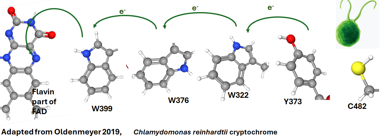
This paper also used the C482A and extended C-terminus mutant. Time-resolved Fourier transform infrared spectroscopy was used to scrutinize tyrosine 373 in several variants of CraCry. The flow of electrons is part of the featured image.
Lacombat 2019, Y373, the source of the long lived red?
The cornestone of this metholdology was the comparison of wildtype with Y373F, phenylanalanine, with no charged residues.
Oxidation of Y373 by coupled electron transport to WH●+ and deprotonation then proceeds in ~800 ps, without any significant kinetic isotope effect, nor a
pH effect between pH 6.5 and 9.0. The FAD●‒/Y373● pair is formed with high quantum yield (~60%); its intrinsic decay by recombination is slow (~50 ms), favoring reduction of Y373● by extrinsic agents and protonation of FAD●‒ to form the long-lived, red-light absorbing FADH● species. Possible mechanisms of tyrosine oxidation by ultrafast proton-coupled ET in CraCRY, a process about 40 times faster than thearchetypal tyrosine-Z oxidation in photosystem II, are discussed in detail.

- Light-induced FADH● formation in CraCRY ultrafast reduction of excited FAD to FAD●‒ by the proximal tryptophan (0.4 ps) WH●+ radical along the tryptophan triad (3.7 and 55 ps). W is the single letter code for tryptophan.
- Oxidation of Y373 by coupled electron transfer to WH●+ deprotonation proceeds in ~800 ps, Y is the single letter code for tyrosine.
- The FAD●‒/Y373● pair is formed with high quantum yield (~60%)
- slow recombination (~50 ms), favoring Y373●
- protonation of FAD●‒ yielding long-lived, red-light absorbing FADH● species.
Red region absorbance between 600-650 nm was mentioned for FADH● in this manuscript. Is FADH located in similar positions relative to tryptophan triads and such?
Tyr373 in CraCry is part of a tetrad of aromatic amino acids see featured image. Subsequently, a nearby cysteine residue at position 482 is oxidized with low yield to form a disulfide bridge, leading to a partial tetramerization of the dimer. TyrO⋅ was suggested to play a key role in the signal transduction of CraCry.


C384 is very close to Y373 which is hydrogen bonded to M486 in the crystal structure. Also note that C482 is hydrogen bonding to many residues that are not tyrosines. Are these interactions even the same in an environment outside a protein crystal? The very interesting thing is that C482 is H-bonded to D371, an amino acid (D387.side chain important human Cry2.
Che 2015, Cry2-CIB1 complexes, a human Cry2 end game?
Human Cry2 lacks the CTT cysteine that is thought to be required for CraCry dimerization and transcriptional activation. These authors used a light insensitive light-insensitive mutant CRY2(D387A). Given the many Cry2 binding partners listed on UniProt, it would be grossly presumptuous to think there is one endgame for Cry2 photo and/or magnetic activation. The Calcium Integrin Binding Protein 1 (CIB1) is a particularly intriguing even though human Cry2 likes an equivalent of C482, it has plenty more in its C-terminus. Che 2015 cited previous studies showing blue light oligomerization of Cry2. CIB1 was tagged with a green fluorescence to monitor cluster formation. Other signaling enzymes were added to the CIB1-GFP complex. While CRY2 homo-oligomerization and CRY2-CIB1 heterodimerization are not mutually exclusive, the presence of certain CIB1 fusion proteins can suppress CRY2 homo-oligomerization. The homo-oligomerization of cytoplasmic CRY2 can be significantly intensified by its recruitment to the membrane via interaction with the membrane-bound CIB1. D387 seems to play a role in this oligomerization. Some of this clustering seems to occur on teh endoplasmic reticulum membrane in COS7, #3T3, and HEK293 cells. Fig 7 of this publication showed similar blue light oligomerization of CRY2 and CIB1 by membrane anchoring Cry2, and attaching green fluorescence protein to Cry2 instead of CIB1. Fig Their cartoons will not be repeated on this post. Instead a cryo electron microscope structure of the plant Arabidopsis thaliana Cry2 tetramer with two CIB1 fragments will be presented:

T476 of Cry2 is on the periphery of these alpha carbon traces. More information on this structure can be found in Zhang 2022.
Ground state oxidized FAD crytochromes are blue light absorbing proteins. The caveat is that unusual amino acid substitutions can prolong red light absorbing semi-quinone states. Whether PEMF can prolong the semi-quinone states is another matter. The human Cry2-CIB1 adds a new twist as to what the end came of Cry2 activation(s) is/are. What is the role of integrin and calcium?
THIS IS NOT WORKING!
This post has been so hard to write. How can one force red light into a blue light absorbing protein? Red light biophotons are easy to explain compared to blue light biophotons. If mysterious biophotons were helping PEMF activate mammalian crytochrome, this should not be so hard. This is not to say blue biophotons do not exist. I just couldn’t find good evidence of them.
References
- Beel B, Prager K, Spexard M, Sasso S, Weiss D, Müller N, Heinnickel M, Dewez D, Ikoma D, Grossman AR, Kottke T, Mittag M. A flavin binding cryptochrome photoreceptor responds to both blue and red light in Chlamydomonas reinhardtii. Plant Cell. 2012 Jul;24(7):2992-3008. PMC free paper
- Bókkon I, Salari V, Tuszynski JA, Antal I. Estimation of the number of biophotons involved in the visual perception of a single-object image: biophoton intensity can be considerably higher inside cells than outside. J Photochem Photobiol B. 2010 Sep 2;100(3):160-6. Sci-Hub free paper
- Che DL, Duan L, Zhang K, Cui B. The Dual Characteristics of Light-Induced Cryptochrome 2, Homo-oligomerization and Heterodimerization, for Optogenetic Manipulation in Mammalian Cells. ACS Synth Biol. 2015 Oct 16;4(10):1124-35. PMC free paper
- Mould RR, Mackenzie AM, Kalampouka I, Nunn AVW, Thomas EL, Bell JD, Botchway SW. Ultra weak photon emission-a brief review. Front Physiol. 2024 Feb 14;15:1348915. doi: 10.3389/fphys.2024.1348915. PMC free paper
- Kar RK, Miller A-F, Mroginski M-A Understanding flavin electronic structure and spectra WIRES Computational Moleculear Science Volume12, Issue2 March/April 2022 e1541 free paper
- Lacombat F, Espagne A, Dozova N, Plaza P, Müller P, Brettel K, Franz-Badur S, Essen LO. Ultrafast Oxidation of a Tyrosine by Proton-Coupled Electron Transfer Promotes Light Activation of an Animal-like Cryptochrome. J Am Chem Soc. 2019 Aug 28;141(34):13394-13409. free paper
- Oldemeyer S, Franz S, Wenzel S, Essen LO, Mittag M, Kottke T. Essential Role of an Unusually Long-lived Tyrosyl Radical in the Response to Red Light of the Animal-like Cryptochrome aCRY. J Biol Chem. 2016 Jul 1;291(27):14062-14071. PMC free paper
- Oldemeyer S, Mittag M, Kottke T. Time-Resolved Infrared and Visible Spectroscopy on Cryptochrome aCRY: Basis for Red Light Reception. Biophys J. 2019 Aug 6;117(3):490-499. PMC free paper
- Schwinn K, Ferré N, Huix-Rotllant M. UV-visible absorption spectrum of FAD and its reduced forms embedded in a cryptochrome protein. Phys Chem Chem Phys. 2020 Jun 10;22(22):12447-12455. free paper
- Usselman RJ, Hill I, Singel DJ, Martino CF. (2014) Spin biochemistry modulates reactive oxygen species (ROS) production by radio frequency magnetic fields. PLoS One. 2014 Mar 28;9(3):e93065. free paper
- Wang J, Liu Y. Systematic Theoretical Study on the pH-Dependent Absorption and Fluorescence Spectra of Flavins. Molecules. 2023 Apr 8;28(8):3315. PMC free paper
- Zhang L, Malkemper EP. Cryptochromes in mammals: a magnetoreception misconception? Front Physiol. 2023 Aug 21;14:1250798. PMC free paper
Leave a Reply