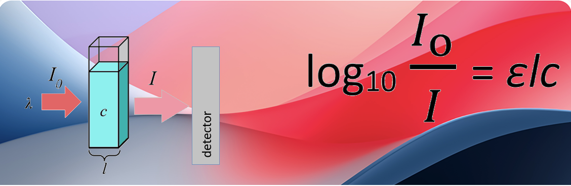
This post is a sister post to red light for depression, which asserts that red and NIR light help alleviate depression by increasing blood flow and functional connectivity. This post focuses on a theoretical study of Bhattacharya & Dutta 2019 exploring what it means to absorb red and near infrared (NIR) light. These authors focus on water, fat, hemoglobin, and cytochrome C oxidase. When light is absorbed by any molecule, that energy can undergo vibrational decay, thus heating nearby molecules. Bhattacharya & Dutta 2019 leaves the impression that there are massive amounts of cytochrome C oxidase in our brains. Does this mean that red light revs up ATP production in our brains? This post will instead try to give the lay reader a refresher course on absorption of light and they can decide for themselves it transcranial red light is all about cytochrome C oxidase. At least we do not have to worry about near infrared heating our brains up.
The science of light absorption
Bhattacharya & Dutta 2019 is not a lay friendly theoretical study. I think it is a lovely, thought provoking study that deserves its own post to go over the wonder of each figure. The featured image illustrates what some of us learned in school as the Beer-Lambert Law. Scientists calculate the concentrations of solutions by placing those solutions in clear, thin tubes. Light is passed through the tube in the solvent used to dissolve the compound. The spectrophotometer measures the amount of light that comes through. This is considered “zero”. The more stuff in that little tube (see featured image) that absorbs light I0, the less light ( I in the image) hits the detector.
ε, a number that tells us how well a compound absorbs light
ε is a constant called the Extinction coefficient or sometimes the molar absorptivity coefficient. The bigger it is, the more light a given compound absorbs per unit concentration, per path length.
In the laboratory a dissolved compound is placed in a square shaped tube 1 cm in diameter. The amount of light that comes through is less. The amount of light of a given wavelength that is absorbed depends on the concentration and the length of the path.
How scientists use ε
Make note of the featured image. ε is a constant with the right units to cancel out all of the units of concentration (usually in moles) and the length (usually in cm). ε is usually in units of per cm and per molar (cm-1M-1). If we know he amount of light absorbed and the path length, we can calculate the concentration. The bigger ε is, the the more light of a particular wavelength the compound can absorb.

Oxy (HbO2) and deoxy (Hb) hemoglobin absorb in the red, near(NIR), and far (FIR) infrared. The X-axis is the wavelength of light that went through the cuvette. Normally the Y-axis is the absorbance. The concentration is calculated at a peak absorption, like 760 nm using a ε specific to that wavelength.
The Y-axis is ε in units of per moles per liter (M-1) per 1 cm (cm-1) The take home is that ε tells us how well a substance absorbs a specific wavelength of light.
- Oxy hemoglobin (HbO2) absorbs more strongly at 840nm and probably at 1064 nm
- Truong 2022 used the for Hb and HbO2 and measured the absorbances of specific wavelengths of red light. In this way they calculated the concentrations of HbO2 to infer blood flow and functional connectivity.
.Xu 2018 proposed that the appropriate wavelength in the 800-1060nm range is good at kicking carbon monoxide or nitric oxide off hemoglobin.
Now that we have an idea of what ε tells us, let’s look at some ε for components of our brains according to Bhattacharya & Dutta.
Overview of Bhattacharya & Dutta figures, with comments
These figures are probably copyrighted. Click on the link and acquaint yourself with what the images are trying to say. Scientists often go directly to the figures of a publication first to engage that part of their brains. The post strives to be such a guide.
Fig 1 Scalp, bone, white, and gray matter absorb red and NIR light
There are many things in our brains that can absorb red light: blood, cerebral spinal fluid, gray matter, white matter… Also note that the paths that red light must travel are greater than just 1 cm. Section 5.2 of Wang 2018 cites research by Jagdeo et al. using human cadaver heads. The authors transmission of 830 nm light: 0.9% through the temporal, 2.1% through the frontal area, and 11.7% through the occipital area.
Table 2 Light can be absorbed and/or scattered
The skull and white matter are close in terms of absorbing 810 nm red light. White matter scatters 830 nm redlight 5x as much as the skull and scalp?! Section 5.2.4 Light Sources of Wang 2025 discusses less light scattering by polarized lasers compared to LEDs, however LEDs can cover a greater surface area.
Liu 2022 go into deeper detail of which major blood vessels in the brain are compromised in depression. How deep are these vessels within our brains? How much gray or white matter must the light travel though before reaching these vessels? How much is the tone of these these vessels affected by said gray and white matter?
Table 3 Bone, CSF, brain, and scalp can conduct and generate heat
If red light is heating parts of the brain up, a good blood flow may cool it down. Metabolically active parts of the brain may generate their own heat irrespective of red light. These are just some numbers used in the computer model and a reminder to those of us who irradiate our tissue with red light. Section 5.2.3 Operation Mode of Wang 2025 discusses pulsed vs continuous red light in terms of heating the tissue. Cerebral spinal fluid (CSF) is not metabolically active and therefore not a generator of heat. Both publications bring up heat as enzymes tend to be a bit more active as the temperature increases. In addition to generating heat, Liu 2022 informs us that metabolic activity generates CO2 that deceases pH and gates L-type Ca2+ channels, that Wikipedia authors tell us are involved in excitation/contraction coupling in blood vessels and skeletal muscle.
Just another point, active mitochondria are generators of heat themselves, particularly in the presence of the uncoupler protein. Why we warm-blooded endothermic animals want to heat our bodies is embedded in the Q10 rule which states that for every 10oC temperature increases, reaction rates double. Keeping a stable temperature allows us to think faster, we just want to keep ourselves in a narrow temperature sweet spot.
Table 4 The brain is the biggest generation of heat in our heads
This table addressed concern that red and NIR can heat our brains up, perhaps in a deleterious fashion. The metabolic activity of our brains is a source of heat as is the blood that slows though our scalps, skull, and brains. 4.1 Cerebral Blood Flow of Wang 2025 discusses the role of nitric oxide synthase in cerebral blood flow response to red light. Liu 2022 are very elaborate in their discussion of how reduced blood flow in depression are the result of vascular dysfunction.
Fig 2 H20 absorbs around 780nm NIR.
Wang 2025 have very little to say about water absorption of NIR. Fig 3 includes a 780 nm study showing reduction of neruro-inflammation cytokines. Bhattacharya & Dutta is a kind of fun, thought provoking figure. Take a look at the very small absorption coefficient at 0.03 per cm.
Fig 3 Lipids absorb around 900-960 nm.
Make careful note that the absorption units in this figure are in unis of “per meter.” This would be 0.15 per cm, to reference back to water. Lipids surround the heat sensitive TRPC1 Ca2+ that is thought to be a target of red light and known to be a target of the capsaicin we use for aches and pains. ε peaks around 15 in this region. Wang 2025 Fig 2 includes TRPC1 as one of many red light depression mechanisms. The lipid peak of 940 nm is interesting and asks the question of fine tuning NIR therapy for enzymes that are associated with lipids. And we get back to he Q10 rule stuff and whether absorbing NIR heats things up just a tad in a very lipid restricted manner. This is something we do not know one way or another. The Wang 2025 review cites 908 nm as being the activator of the TRPV1heat/capsaicin receptor. TRPV1, a non specific Ca2+ channel, is also a receptor for heat and CBD. Wikipedia authors have written on its desensitization with agonists for pain relief. TRPC1 and TRPV1 are members of the same “Transient Receptor Potential” Ca2+ channels.
Fig 4 Cytochrome C oxidase is an absorber of red light
CCO is named because it takes an electron from cytochrome C, thereby oxidizing it. In this process, it becomes reduced. Once t ercieved this electron, it transfers it to molecular oxygen reducing it to water. In this process, CCO becomes oxidized. Cytochrome C oxidase, reduced and oxidized, absorb in the blue range and at 600nm. Only the oxidized absorbs 800 nm red light well. What does this mean if we ant to use red light to increase flux of electrons through complex IV of the electron transport chain? What does this mean in terms of pumping H+ out that are used to make ATP? Wang 2025 have a section on cytochrome C oxidase but do not report on directionality. They do point out that red light can release nitric oxide from cytochrome C oxidase allowing flow of electrons. Nitric oxide has more important action in increasing blood flow covered in the spinoff post.
Fig 5 Blood absorbs red light
Oxy and deoxy hemoglobin absorption: 600-700 nm range. HbO2 absorbs well in this range and deoxy Hb does not. This is a fine and frustrating figure in that nitroso hemoglobin is not included. Luchsinger 2003 is a study about hemoglobin as a carrier of nitric oxide. Fig 1 shows visible light absorption spectra for met hemogobin (green diamonds), Fe (II)NO (red circles), and Fe(III) (black squares) Unfortunately this figure does not extend into the NIR range.
Table 5 2 heme groups, 2 oxidation states
This table tells us that how much of any of these red light absorbing compounds depends on how much there is in any one part of the brain. Deoxy hemoglobin and oxidized cytochrome C oxidase seem to be big sinks for 630nm red light in gray matter. Just look at the difference in molar absorption coefficients that tell us where the red and NIR might be acting. This is a very interesting gray vs white matter computer experiment. White matter has more myelination, hence fat. Cytochrome C oxidase oxidizes cytochrome C then it reduces O2 to H20… The O2 comes from HbO2… Can we fine tune this process by fine tuning the wavelength of red and NIR light?
Table 6 whole brain tissue absorption
Never mind hemoblobin and cytochrome C oxidase, this able compares whole tissue gray and white matter at 630, 700, and 810 nm. Gray matter absorbs twice as much red light wave lengths. This post is going to drop the rest of the very thought producing narrative of Bhattacharya & Dutta 2019 because Weerasekera 2024 demonstrated no global brain heating with red light therapy. This post maintains that other heme containing enzymes are probably as important as cytochrome C oxidase and hemoglobin that the clinician should be aware of.
Liu 2024 made a case for neuronal activity regulating blood flow. This is where nNOS comes in even though Truong 2022 didn’t conclude it was that important in the brains’ response to 1064 nm NIR!
References
- Bhattacharya, M., & Dutta, A. (2019).Computational Modeling of the Photon Transport, Tissue Heating, and Cytochrome C Oxidase Absorption during Transcranial Near-Infrared Stimulation. Brain sciences, 9(8), 179. PMC free article
- Keszler A, Lindemer B, Hogg N, Lohr NL. Ascorbate attenuates red light mediated vasodilation: Potential role of S-nitrosothiols. Redox Biol. 2019 Jan;20:13-18. PMC free paper
- Liu M, He E, Fu X, Gong S, Han Y, Deng F. Cerebral blood flow self-regulation in depression. J Affect Disord. 2022 Apr 1;302:324-331. free paper Blood flow is mucked up in depression.
- Truong NCD, Wang X, Wanniarachchi H, Liu H. Enhancement of Frequency-Specific Hemodynamic Power and Functional Connectivity by Transcranial Photobiomodulation in Healthy Humans. Front Neurosci. 2022 Jun 10;16:896502. PMC free paper
- Wang L, Mao L, Huang Z, Switzer JA, Hess DC, Zhang Q. Photobiomodulation: shining a light on depression. Theranostics. 2025 Jan 1;15(2):362-383. PMC free paper This is a very extensive review that can become a bit overwhelming.
- Weerasekera A, Coelho DRA, Ratai EM, Collins KA, Puerto AMH, De Taboada L, Gersten MB, Clancy JA, Hoptman MJ, Irvin MK, Sparpana AM, Sullivan EF, Song X, Adib A, Cassano P, Iosifescu DV. (2024) Dose-dependent effects of transcranial phoobiomodultion on brain temperature in patients with major depressive disorder: a spectroscopy study. Lasers Med Sci. 2024 Oct 7;39(1):249. PubMed
- https://redlighttherapyhome.com/blogs/news/red-light-therapy-for-anxiety-and-depression
“How Red Light Therapy Works for Mental Health
Light is a form of energy. While certain types, like blue and ultraviolet (UV) light, can be harmful in excess, red and near-infrared light have demonstrated numerous therapeutic benefits. This process, known as photobiomodulation, involves exposing the body to specific wavelengths of light to stimulate biological processes.
The human body’s cells contain mitochondria, which are essential for producing cellular energy. When exposed to red or near-infrared light within the therapeutic window of 600 nm to 1000 nm, these mitochondria are activated. This stimulation can lead to increased cellular energy, growth, and repair.
Initially, researchers believed these benefits were primarily physical. However, emerging research suggests that photobiomodulation may also be applied to support mental health, with studies exploring its effects on conditions like PTSD, depression, and anxiety. While questions remain about the depth of light penetration required for neurological conditions like Parkinson’s or Alzheimer’s disease, the therapy shows potential for influencing brain states related to mood.
It is important to note that red light therapy should be considered an adjunctive treatment and not a replacement for professional mental health care or established therapies.”
Red light therapy Home RLTH favors a more multimodal explanation of how red and NIR light improve our mental health. It’s hard to tell which mode comes first. The theoretical study of Bhattacharya & Dutta 2019 certainly make it seem like it is all about cytochrome C oxidase. Life is seldom that simple. Even if red/NIR light is all about CCO, one thing that Bhattacharya & Dutta 2019 did not show us was the absorption spectrum of nitrosylated CCO. Does one wavelength of red/NIR kick the NO group off better than another? Kicking nitric oxide off heme irons and protein thiols as a means of increasing blood flow was addressed in the to red light for depression post.
If you would like to order your own red light therapy device from Red Light Therapy Home, use this promo code for a 6% discount
Leave a Reply