This post takes the model of Franco-Obregon 2023 that the QuantumTx brand of PEMF gates calcium currents of the transient receptor potential (TRP) family member TRPC1 by way of producing reactive oxygen species that modify the TRPC1 channel. There are some cysteines near some regulatory phosphorylation sites, calcium calmodulin binding sites, and ankyrin repeats, structures with unknown binding partners in TRPC1. As far as I know .
Domains of TRPC1
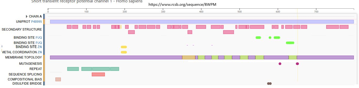
The featured image is from rcsb.org, the crystal structure of one TRPC1 in a tetramer with three TRPC4 subunits. It takes four monomers to form a channel. TRPC1 does not form homotetramers. 8WPM TRPC1 is close to 800 amino acids in length
- Secondary structure: The first row appears to be alpha helices and the second beta sheets
- Unmodeled is stuff that is flopping around in the breeze too much to be visible in the crystal structure
- PJQ is a ligand. I’m not sure if it is an agonist or antagonist of calcium
- We’ve got Ca2+ binding sites and a site where Mg2+ maintains the protein structure but does not transported.
- Membrane topology is the parts of the protein that go in the cell membrane.
- Mutagenesis, two amino acids, when mutated, increase or decrease the flux of Ca2+ through the channel.
- The repeats are ankryin repeats.
- Sequence splicing occurs on the mRNA level and reflects a shorter isoform of the channel.
- And of course, disulfide bonds involve cysteines that will not be subject to oxidation by cryptochrome derived reactive oxygen species.
Detecting magnetic fields
Franco-Obregón 2023, Figure 3 Click here to view an impressive cartoon showing Hypothetical model of magneto reception and the factors that may influence its operation. Orange circles in Fig 3 represent Ca2+ ions and red circles represent ROS. Reactive oxygen species and environmental factors such as photons, temperature, and pH are proposed to be part of the process. Figure 10 of Wiltschko 2014 of Wiltschko 2014 models how visible light and magnetic fields work in cryptochrome if avian
A look at post translational modifications of TRPC1 Liu 2020
Domains and post translational modification of TRPC family have been discussed extensively in an excellent review. Liu 2020. Some of this image were adapted from Liu 2020 including the phosphorylation sites that negatively gate Ca2+ currents. Singh 2002 demonstrated two CaM binding domains on the TrpC1-C terminus, aa 719–751 and aa 758–793.

This cartoon started out loosely based on Liu 2020. Ankyrin repeats were changed based on the motif of two alpha helices separated by a small linker. Some cysteines were found in these motifs. Wikipedia has little to say on ankyrin repeats save they may be solenoid in shape and areas of signal transduction. I’ve tried to render a partial solenoid There’s also a list of proteins that have these repeats including TRP family members. The three phosphorylation sites are from Liu 2020. Note that there are five cysteines around these phosphorylation sites! Cysteine sulfinic and sulfonic acids could theoretically mimic phosphorylation. The 346-375 cylinder is a pre-transmembrane alpha-helix. It spans some cysteines. There are no transmembrane cysteines. There’s a disulfide bond and one reduced in the extracellular loop that folds into the channel mouth. The actual channel has three additional subunits, e.g. three of TRPC4 monomers.
Ca2+calmodulin binding sites are also found in TRPC4 that forms hetero tetramers with TRPC1. (Chen 2020) All well and good. Are the cysteines close to the CaCam site conserved and regulatory? If so, what is the consequence of oxidation to cysteine modification that carries a charge?
We’ve got a few cysteines close to the ankyrin repeats. A search was conducted to find the binding partners of these ankyrin repeats. There isn’t just one. These flexible structures have the potential to have antibody like specificity. Designed Ankyrin Repeat proteins (DARPin) have their own Wikipedia page. To the best of my knowledge, no one has ever tried to use expressed ankyrin repeats of various TRP channels as proteomics bait in an effort to determine if these repeats have the capacity to bind to different effector proteins. Perhaps the repeats of TRPC1 selectively bind to cryptochrome compared to the ankyrin repeats of other TRP family members or other proteins altogether.
References
- Chen X, Sooch G, Demaree IS, White FA, Obukhov AG. Transient Receptor Potential Canonical (TRPC) Channels: Then and Now. Cells. 2020 Aug 28;9(9):1983 PMC free paper
- Franco-Obregón A. Harmonizing Magnetic Mitohormetic Regenerative Strategies: Developmental Implications of a Calcium-Mitochondrial Axis Invoked by Magnetic Field Exposure. Bioengineering (Basel). 2023 Oct 10;10(10):1176. PMC free article
- Liu X, Yao X, Tsang SY. Post-Translational Modification and Natural Mutation of TRPC Channels. Cells. 2020 Jan 7;9(1):135 PMC free article
- Singh BB, Liu X, Tang J, Zhu MX, Ambudkar IS. Calmodulin regulates Ca(2+)-dependent feedback inhibition of store-operated Ca(2+) influx by interaction with a site in the C terminus of TrpC1. Mol Cell. 2002 Apr;9(4):739-50. free paper
- Wiltschko R, Wiltschko W. Sensing magnetic directions in birds: radical pair processes involving cryptochrome. Biosensors (Basel). 2014 Jul 24;4(3):221-42. PMC free article
Leave a Reply