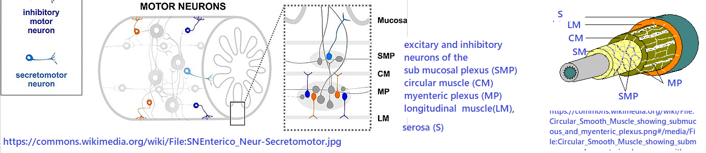
The featured image, the enteric nervous system
The featured image is a Wikicommons image of the enteric nervous system. Here’s an okay YouTube video on peristalsis, the moving of bolus through tubes in our bodies. The colon and small intestines have two layers of smooth muscle that take turns contracting and relaxing. According to this YouTube video, the stomach contains three layers of smooth muscle to accommodate mixing of food during the first stage of digestion. This post takes me back to my early twenties as a tech in Dr Don Powell’s lab for a short while. This is what I saw each time I killed a rat for its colon. Even after the colon has been removed, the pellets continue to move “south.”

Even back in the day the enteric nervous system was called the “Little Brain.” The thought process was that the cells of the myenteric and submucosa plexus could be exposed to cells of the immune system that secrete cytokines that control how well the epithelial cells transport electrolytes, and hence the water content of the pellets. The myenteric plexus, between circular and longitudinal smooth muscle, that must work together to move pellets. Imagine the agony a horse must feel when they too experience pellets that just get stuck. How lucky are we humans that our appendix is nothing like the cecum of a rat, horse, or rabbit.
Colic in Horses, causes
Horse colic has it’s own Wikipedia page. It became necessary to consult the ordinal reference: Abutarbush SM, Carmalt JL, Shoemaker RW. Causes of gastrointestinal colic in horses in western Canada: 604 cases (1992 to 2002). Can Vet J. 2005 Sep;46(9):800-5. PMC free paper.
| Diagnosis | explanation | Percentage |
|---|---|---|
| Large colon impaction | pellet gets stuck | 20.8 |
| Large colon displacement | colon moved | 16.5 |
| Spasmodic colic | very painful GI contractions | 11.7 |
| Large colon volvulus | twisted, loop of intestine | 7.3 |
| Lipoma | benign fat tumor | 6.9 |
| Strangulating small intestine lesion (other) | cuts off blood supply, could be lipoma | 4.2 |
| Enteritis | small gut inflammation | 3.4 |
| Peritonitis | may involve GI perforation | 2.7 |
| Verminous arteritis a worm | mesenteric parasite | 2.1 |
Dr Robert N. Oglesby DVM covers much of this in a review on HorseAdvice.com. The interesting thing to note is that just like the rat colons I have observed, pellets getting stuck is a leading cause of “colic” in horses. Percentage wise, movement of the colon was a close 2nd. Human intestines are resting on the pelvic floor. The intestines of our four legged companions rest on abdominal muscles. The volumes of chyme/pellets in the large and small colon can easily be equal in weight to a male human rider. This is on top of the volume of material in the stomach and cecum!
The volume of GI compartments in horses
Here is an image is an adaptation from this site Volume in quarts have been converted to liters for easy conversion to kg.

MagnaWave narrative
The following has clearly been written by a DVM or another trained person who knows a lot about horses.
“Colic affects between 4 and 10 percent of horses during their lifetimes. It is a serious and sometimes life-threatening condition. It should be noted that colic surgery is still a risk for horses.
Horses that suffer through Colic have not had many treatment options in the past, but now there is a new option. MagnaWave uses PEMF technology to help reduce your horse’s pain and get them “going” again!
According to the American College of Veterinary Surgeons, a few symptoms you might notice if your horse is suffering from colic may include:
- Depression
- Inappetence (not interested in eating)
- Pawing
- Looking at the flank
- Lying down more than usual or at a different time from normal
- Lying down, getting up, circling, laying down again repeatedly
- Curling/lifting the upper lip
- Kicking up at the abdomen with hind legs
MagnaWave has been used many years for horses by using PEMF technology to reduce pain and inflammation and hasten healing. PEMF works by producing a cascade of biological processes that support ailing tissues. There has been limited research completed on PEMF for colic in horses. Veterinarians use results from research done on other species to determine what will work best to treat colic in horses. The horse’s physical response to PEMF allows the veterinarian to know whether it is having a positive effect.”
Gastric Ulcers in Horses
Helping Gastric Ulcers in Horses Naturally with MagnaWave. Much of the cause of gastric ulcers is fact checked on Horseadvice.com.
“Gastric ulcers are commonly found in horses. A third of adult horses confined in stalls and over 60% of performance horses develop moderate to severe gastric ulcers.
Horses constantly produce hydrochloric acid which can accumulate in the stomach when the horse does not eat often enough. This causes an acid build-up that can lead to gastric ulcers. With gastric ulcers being such a common occurrence, owners and trainers are MagnaWaving gastric ulcers in horses naturally with PEMF.
MagnaWave with Gastric Ulcers
PEMF uses pulses of electromagnetic waves deep into damaged and diseased tissue. The coils can be strategically placed and emit a measurable magnetic field up to 16 inches. This electromagnetic field helps cells to function normally and strengthens cell metabolism.
MagnaWave uses PEMF to helps to reduce inflammation and promote healing. It can be used as an adjunct modality to help the horse feel better as other changes in management are made.
Addressing gastric ulcers in horses naturally with PEMF is safe, non-invasive and has no adverse side effects. It requires no sedation and is both calming and relaxing to the horse.”
⚠️ my cautionary note
There is no way I am going to “MagnaWave any equine without the supervision of a veterinarian. For starters, I’ve no history of interacting with any equine species. To continue, I know way too much of possible dangers if the source of colic is unknown. I have seen way too many impacted, convoluted, and spasmodic rat colons to even remotely consider addressing such issues on a much larger scale. The obvious vet influence of the MagnaWave narrative fills me with curiosity as to how this technology might work with colicky canines, felines, or even humans. Those memories of rat colons prevents me from working outside the collaboration of someone who is trained to practice medicine.
TRPC1 in the myenteric plexus
Liu S, Qu MH, Ren W, Hu HZ, Gao N, Wang GD, Wang XY, Fei G, Zuo F, Xia Y, Wood JD. Differential expression of canonical (classical) transient receptor potential channels in guinea pig enteric nervous system. J Comp Neurol. 2008 Dec 20;511(6):847-62. PMC free article
This particular publication looked a TRPC family members 1-7. PEMF “antenna” TRPC1 is of particular interest to this site.
- TRPC1-IR was restricted to nerve cell bodies and proximal processes and not found in varicose nerve fibers.
- TRPC1- human antibody immuno reactive nerve cell bodies accounted for 63.2 ± 7.8% of the total myenteric neuronal population.
- In the myenteric plexus TRPC1 was colocalized with choline acetyltransferase, calretinin, nitric oxide synthase, but not calbindin
- In the submucosal plexus, TRPC1-immuno reactive neurons accounted for 67.7 ± 11.6% of the total submucosal neuronal population.
- In the submucosal plexus TRPC1 colocalized with choline acetyl transferase, calretinin, neuropeptide Y, and vasoactive intestinal peptide, but not calbindin or neuronal nuclear protein.
In summary, a Ca2+ channel that has been described as an antenna for PEMF is colocalized with with enzymes like nitric oxide synthase that can lead to smooth muscle relaxation.
Voltage gated Ca2+ channels in the myenteric plexus
PEMF and VGCC (voltage gated Ca2+ channels) has been addressed on this site. A through review of all the subtypes of CGCC and whether or not they respond to PEMMF has not been conducted.
Reis HJ, Bíscaro FV, Gomez MV, Romano-Silva MA. Depolarization-evoked GABA release from myenteric plexus is partially coupled to L-, N-, and P/Q-type calcium channels. Cell Mol Neurobiol. 2002 Dec;22(5-6):805-11. PubMed
This study found almost no influence of L- and Q-type calcium channels, while N- and P-type only contributed 15% of the GABA release. The authors speculated that the intracellular Ca2+ increase needed for GABA release probably came from intracellular stores. TRPC family members are couple to intracellular Ca2+ stores.
Leave a Reply