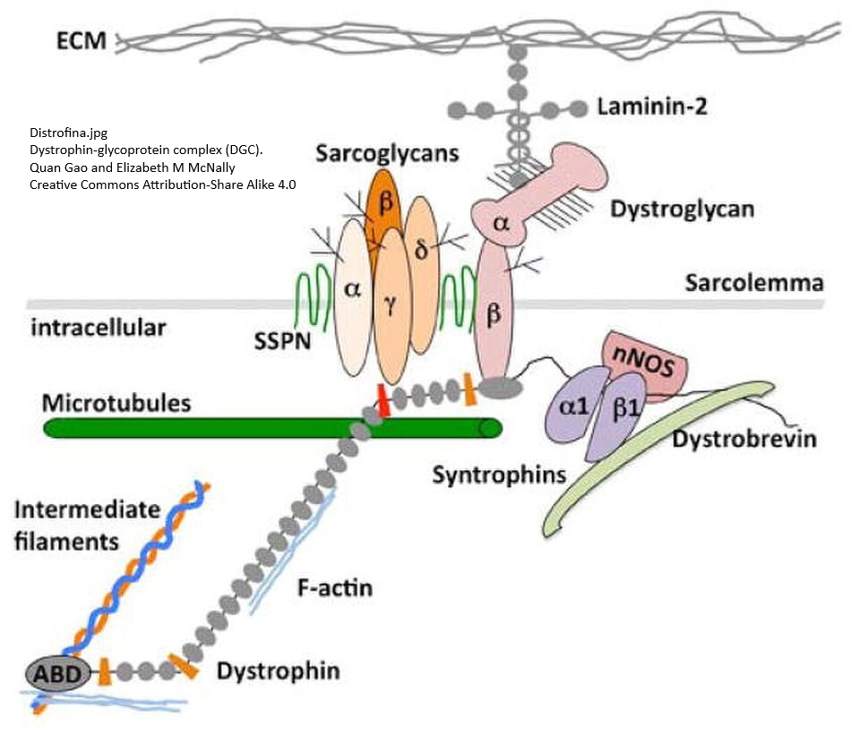
Can PEMF reverse vascular aging related to decreased protein levels of dystrophin, what is commonly thought of as a skeletal muscle protein mutated in Duchene and Becker muscular dystrophies. This link will take you to a cartoon showing subtle differences between dystrophin and its more ubiquitous homolog utrophin. The two molecules are very similar with some subtle differences. Dystrophin is absent in two forms of muscular dystrophy. Iversen and Franco-Obregon published a review/proposal that PEMF could convince skeletal muscle to produce the homolog utrophin when the gene for dystrophin is faulty. The mechanism involves gentle activation of TRPC1 Ca2+ channels and activation of two Ca2+ transcription factors. This post also examines a paper that mapped the utrophin promoter and added just a little to the mitohormesis them of the Iverson & Franco-Obregon review.

Urophin, on the other hand, is everywhere. We don’t know how hard the scientists who compiled these data were looking. I think the most striking thing is the expression in endothelial cells that line our blood vessels. It is the endothelial cells that respond to shear forces of blood flow. Iverson and Franco-Obregon 2026 make a case for using PEMF as a kind and gentle way of boosting utrophin expression in skeletal muscle to compensate for defective dystrophin. Perhaps it can be a kind and gentle way of boosting utrophin expression elsewhere.

Iversen and Franco-Obregón 2026, a review and a proposal
QuantumTx is a Singapore based company with a primary market focus of helping senior citizens age with muscular cellular quality of life via a process that they call “Mitohormesis.” Hormesis means that things that are toxic in large amounts can be health promoting in small amounts. Small amounts of superoxide can activate the TRPC1 Ca2+ channel.
Fig 1 is probably copyrighted. View the figure in a different tab.
(A) Glycolytic muscle fibers:
At rest
Unlike the featured image, we’ve got the ghost of dystrophin tugging at some Ca2+ TRP channels. TRPC1 is the main one. The graphical abstract of Le 2018 is a beautiful cartoon of the folding and unfolding of domains of dystrophin.
Under mechanical stress
Sarcolemmal rupture and dysregulated TRPC1/3 activity cause excessive Ca2+ influx, high ROS, and actin degeneration as a result of the dystrophin shock absorbers. α1-syntrophin is still attached to TRPC1, but there is no mechanotransduction. The TRPC1 Ca2+ channel is forming a heterotetramer with its cousin Ca2+ channel TRPC3 that activates NADPH oxidase (Nox2) and free radical production. The review/proposal covered physical interactions of TRPC3 and Nox2
PEMF stimulation
PEMF stimulation may trigger non-mechanical TRPC1-mediated Ca2+ entry, activating calcineurin–NFAT signaling to upregulate utrophin and induce UGC compensation.
Utrophin is a dystrophin associated protein that has a fairly high degree of homology to dystrophin. Dystrophin and Utrophin can form Vulnerable phenotype characterized by low mitochondrial density and basal utrophin expression. Resilient phenotype characterized by high mitochondrial density and basal utrophin expression. Preserved membrane stability maintains UGC–TRPC1 tethering, limiting pathological Ca2+ entry and ROS during mechanical stress. PEMF enhances this protective profile by reinforcing UGC stability and oxidative capacity. The take home seems to be that even though utrophin is connected to slightly different membrane proteins, it still has the capacity to dampen some of the mechanical strain of contraction by way of its spring like domains.
According to the Noize 2023 review, calcineurin–NFAT is very much part of vascular smooth muscle dignaling, responding to and increasing the expression of TRPC Ca2+ channels. Utrophin and dystrophin were not mentioned.
(B) Oxidative muscle fibers:
“Oxidative muscles also exhibit the highest expression levels of TRPC1, where it promotes oxidative (type I) muscle development. By contrast, the expression of TRPC3 is induced by mechanical forces in both fast glycolytic (type II) and slow oxidative (type I) muscle, and its expression is often inversely correlated to that of TRPC1.”
(C) Signalling mechanism and phenotypic remodelling:
Panel C is the cornerstone of QuantumTx technology, that PEMF also induces the transcription of mitochondrial genes on the chromosome. The transcription of a dystrophin homolog is a new twist. Increasing transcription of dystrophin in aging muscles would also be in interesting twist to the story.
Dystrophin deficiency in vascular smooth muscle Xuan 2025
findings suggest that dystrophin deficiency drives VSMC phenotype switching and mitochondrial dysfunction, contributing to vascular pathology in DMD. These findings highlight the importance of targeting vascular abnormalities in therapeutic strategies to slow disease progression
- Fig1 Dsytrophin is expressed in multiple types of human smooth muscle and also multiple cell types within that muscle. This figure strongly supports the ProteinAtlas mRNA transcript figure in this post.
- Fig2, There is more fibrosis in the aorta of Mdx mice. Why is this? From what ProteinAtlas has to say, “mural cells” also express utrophin.
- Fig3, TGF-β1 was used to induce the differentiation pluripotent stem cells into vascular smooth muscle cells. VSMCs markers including α-actin (smooth muscle), SM-22α, and calponin were decreased in DMD cells.
- Fig 4 Vascular smooth muscle cell mitochondria from DMD mice are more likely to be fragmented and producing reactive oxygen species. In the desmin mitohormesis post on this site, it was covered that the intermediate filament desmin binds to mitochondria. Is there a connection? Does utrophin bind intermediate filaments like desmin?
- Fig 5 Upregulated and down regulated GO categories of genes. GO 0090280 (Any process that increases the rate, frequency, or extent of the directed movement of calcium ions into a cell or organelle) was about 10x down regulated in DMD differentiated VSMC. We’ve got dystrophin and/utrophin tugging at TRP channels. Are both the utrophin and dystrophin promoters sensitive to Ca2+ pathways?
- Fig6 looked at the response of DMD or WT VSMC to oxidative stress.
Iverson & Franco-Obregon 2026 made a case for PEMF switching dystrophin skeletal muscle from a glycolytic to an oxidative phenotype with more mitochondria. With a reasonable amount of searching I was unable to draw a parallel between tonic and phasic smooth muscle covered in LIbre Texts. One would think that phasic smooth muscle of the gut would have more mitochondria and perhaps even rely on fatty acids as an energy source more, but the text did not say as much. We don’t want our blood vessels contracting like our guts anyway. Compliance is anther matter and another post!
TRP channels in WT vs DMD VSMC, Lopez 2020
“Duchenne muscular dystrophy (DMD) is an irreversible muscle disease characterized by a progressive loss of muscle function, decreased ambulation, and ultimately death as a result of cardiac or respiratory failure. DMD is caused by the lack of dystrophin, a protein that is important for membrane stability and signaling in excitable cells. Although vascular smooth muscle cells (VSMCs) dysfunction occurs in many pathological conditions, little is known about vascular smooth muscle function in DMD. We have previously shown that striated muscle cells, as well as neurons isolated from dystrophic (mdx) mice have higher intracellular Ca2+ ([Ca2+]i) and Na+ ([Na+]i) concentrations and decreased cell viability in comparison with wild type (Wt). Experiments were carried out in isolated VSMCs from mdx (a murine model of DMD) and congenic C57BL/10SnJ Wt mice. We found elevated [Ca2+]i and [Na+]i in VSMCs from mdx mice compared to Wt. Exposure to 1-oleoyl-2-acetyl-sn-glycerol (OAG), a TRPC3 and TRPC6 channel activator, induced a greater elevation of [Ca2+]i and [Na+]i in mdx than Wt VSMCs. The OAG induced increases in [Ca2+]i could be abolished by either removal of extracellular Ca2+ or by SAR7334, a blocker of TRPC3 and TRPC 6 channels in both genotypes. Mdx and Wt VSMCs were susceptible to muscle cell stretch-induced elevations of [Ca2+]i and [Na+]i which was completely inhibited by GsMTx-4, a mechanosensitive ion channel inhibitor. Western blots showed a significant upregulation of TRPC1 -3, -6 proteins in mdx VSMCs compare to age-matched Wt. The lack of dystrophin in mdx VSMCs produced a profound alteration of [Ca2+]i and [Na+]i homeostasis that appears to be mediated by TRPC channels. Moreover, we have been able to demonstrate pharmacologically that the enhanced stretch-induced elevation of intracellular [Ca2+] and concomitant cell damage in mdx VSMCs also appears to be mediated through TRPC1, -3 and -6 channel activation.”
Dystrophin vascular smooth muscle, age Kaplan 2026
“As humans age, the aorta stiffens, diminishing its essential shock absorber function. This increased stiffness transmits higher pressures to downstream vessels in the kidney, brain, and heart, contributing to hypertension and end-organ damage. Although multiple mechanisms involving extracellular matrix (ECM) remodeling and vascular smooth muscle cell (VSMC) contributions to aortic stiffness have been described, additional molecular players likely remain uncharacterized. Dystrophin (DYS) and the dystrophin-associated proteins (DAPs) are cytoskeletal stabilizers known for protecting skeletal muscle cells from contraction-induced damage but are poorly characterized in VSMCs. Loss of DYS has been linked to myocardial stiffness and carotid artery abnormalities. Here, we investigated the function of DYS in biomechanical properties of the mouse aorta. We demonstrated by immunofluorescence that DYS and DAPs are expressed and colocalize in freshly dissociated murine VSMCs. The mdx model, a known dystrophin knockout mouse model, was used to investigate the consequences of lack of dystrophin expression on aortic geometry and biomechanics. In mdx mice, we observed decreased aortic wall thickness but no significant difference in diameter compared to wild-type (WT) mice. This significant difference in aortic geometry is directly related to ex vivo stress and stiffness. We measured aortic stiffness by high-frequency small-amplitude sinusoidal length perturbation and determined dystrophin is essential to maintain normal stiffness and stress at baseline. This suggests that extracellular matrix components in the mdx aorta are contributing to the increased stress and stiffness at baseline. We also determined that DYS is not required to maintain normal stress and stiffness due to the contractile response to depolarization by high K + or alpha-agonist, phenylephrine-induced contraction. Finally, we revealed by Western blot that DYS and alpha-sarcoglycan, a DAP, expression is decreased in aged vascular smooth muscle. Previous studies show that matrix metalloproteinase-2 (MMP2) can degrade DYS in cardiac tissue and that MMP2 activity increases with age in vascular tissue. While we did not directly assess MMP2 in this study, we propose its role as a hypothesis for future exploration. In summary, this study identifies a novel role for DYS in maintaining aortic mechanical integrity and presents evidence that aging diminishes DYS and DAP protein expression in VSMCs.”
This brings us back to the post of a few years ago on PEMF and aorta stiffening in which it was concluded that PEMF probably worked on e level of vascular endothelial cells.
The utrophin promoter
It has been unbelievably hard to find info on the utroppin promoter. Could the same pathway in Fig 1C of Iverson & Franco-Obregon also exist in smooth muscle? Angus 2005 mapped out the utrophin promoter in a way that is entirely consistent with mitohormesis.

GAPBA is a transcription factor that binds to purine (G and A) repeats. In addition to the utrophin gene, GA controls the expression of mitochondrial electron transport chain cytochrome C and cytochrome C oxidase. The Human Protein Atlas was consulted as to whether or not GABPA is found in smooth muscle and “mural” cells which are a combination of smooth muscle and pericytes within tissues.

This post has unfortunately (or fortunately) not gotten into the control of GABPA. Seeing expression of GABPA in metabolically active cells and knowing it targets utrophin expression posses more questions.
References
- Angus LM, Chakkalakal JV, Méjat A, Eibl JK, Bélanger G, Megeney LA, Chin ER, Schaeffer L, Michel RN, Jasmin BJ. Calcineurin-NFAT signaling, together with GABP and peroxisome PGC-1{alpha}, drives utrophin gene expression at the neuromuscular junction. Am J Physiol Cell Physiol. 2005 Oct;289(4):C908-17. PMC free paper
- Kaplan KM, Xiong Y, Lucerne A, Morgan KG. Dystrophin and dystrophin association protein expression decreases with age in vascular smooth muscle. Geroscience. 2026 Aug;48(4):5597-5612. PMC free paper
- Lopez JR, Uryash A, Faury G, Estève E, Adams JA. Contribution of TRPC Channels to Intracellular Ca2 + Dyshomeostasis in Smooth Muscle From mdx Mice. Front Physiol. 2020 Feb 20;11:126. PMC free paper
- Ingueneau C, Huynh-Do U, Marcheix B, Athias A, Gambert P, Nègre-Salvayre A, Salvayre R, Vindis C. TRPC1 is regulated by caveolin-1 and is involved in oxidized LDL-induced apoptosis of vascular smooth muscle cells. J Cell Mol Med. 2009 Aug;13(8B):1620-1631. PMC free paper
- Iversen JN, Franco-Obregón A. Magnetic Mitohormesis as a Potential Non-Invasive Restorative Therapy for X-Linked Muscular Dystrophies. Int J Mol Sci. 2026 Aug 28;27(17):7700. PMC free paper
- Le S, Yu M, Hovan L, Zhao Z, Ervasti J, Yan J. Dystrophin As a Molecular Shock Absorber. ACS Nano. 2018 Dec 26;12(12):12140-12148. PMC free paper
- Nolze A, Matern S, Grossmann C. Calcineurin Is a Universal Regulator of Vessel Function-Focus on Vascular Smooth Muscle Cells. Cells. 2023 Sep 13;12(18):2269. doi: 10.3390/cells12182269. PMID: 37759492; PMCID: PMC10528183.
- Xuan W, Cheng F, Han X, Tipparaju SM, Ashraf M. Impact of dystrophin deficiency on vascular smooth muscle cell. Sci Rep. 2025 Nov 24;15(1):45254. PMC free paper
Leave a Reply