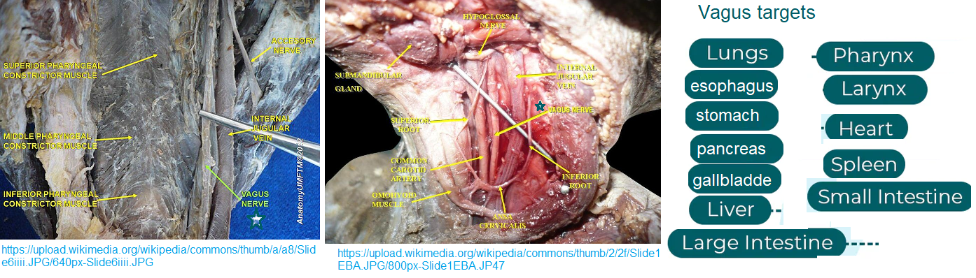
This post takes the Devil’s Advocate position that many of the PEMF devices on the market specifically for vagus nerve stimulation might not be stimulating the nerve directly but rather, hypothetically, clearing away toxins from un-stirred regions around the nerve itself. The featured images of the vagus nerve from a human cadaver bring home how crowded things are. Some vagus nerve targets from proximal to distal bring home the point that stimulating the entire nerve may also stimulate the many branches.
Britanica and Wikipedia have good posts on the vagus nerve. The vagus nerve starts in the brain stem and branches out. The interesting comment on Wikipedia is the large about of afferent information.
An excellent anatomy study of the porcine vagus nerve.
If an electrode is to be placed along the Vagus nerve for stimulation (VNS) we need to know as much as possible about the anatomy of this nerve.
- Given the off-target effects of VNS, there is renewed interest in developing novel stimulation strategies and multi-contact electrodes to stimulate selectively specific fibers within the vagus—typically preganglionic efferents or sensory afferents to and from the visceral organs—while avoiding motor efferent fibers coursing within or near the cervical vagus trunk [1]
- used microdissection and post-mortem histology to quantify anatomical features of the cervical vagus nerve of the domestic pig including nerve diameter, number of fascicles, diameter of fascicles and distance of fascicles from the epineural surface where VNS electrodes are placed. [1]
- SLN of the vagus branches more cranially in humans, the external superior laryngeal (ESL) branch of the superior laryngeal that innervates the cricothyroid muscles can pass near the carotid bifurcation, which in turn can be located quite close to the traditional clinical VNS lead placement (Monfared et al 2001). Consequently, activation of the ESL branch may be a contributor to unwanted neck muscle activation in clinical VNS.
The following are links to histological stains from the publication. The authors used a lot of H&E staining. Purple stains the nuclei and pink lipid and cytosolic regions.
- Image 1 is a cross section of the cervical vagus showing fascicles (bundles) of various nerves. What is the circulation like in these bundles?
- Image 2 of the left and right vagus at the aortic arch and larynx.
- Image 3 the Vagus nerve and carotid artery from two different pigs. One appears to have more fat, the other may have more leaked blood that might have been an artifact of the dissection.
- Image 4 a cartoon of the site of electrode placement for electrical Vagal nerve stimulation.
- Image 5 Depiction of the sympathetic trunk (ST) running parallel to the vagus nerve (VN) in one subject, and also ‘hitch-hiking’ along sections of the nerve including cross-connections to the nodose ganglion (NG); superior laryngeal (SLN), carotid bifurcation (CB), vagus nerve (VN).
- Image 6 number of fascicles, average fascicle diameter, and closest distance from the epineural surface to the nearest fascicle varied greatly across specie
- Image 7 Examples of left and right side vagal nerve cross-sections showing the singular large fascicles of aggregated pseudo-unipolar cell bodies in a single plane when the nodose was sectioned extensively (A), whereas the aggregated pseudo-unipolar cells were not visible in early subjects, when nerves were sampled more sparsely, black arrows indicate a few sparse pseudo-unipolar cells (B).
- Image 8 shows cross sections of the Vagus nerve down the length after numerous bifurcations. The number of fascicles of nerves decreases. Does the microenvironment around a fascicle deep in the fiber differ from one in the process of branching off?
- Image 9 shows Example cross sections of a pig cervical vagus nerve labeled with an antibody against choline acetyltransferase (ChAT), indicated by the brown chromogen (DAB). This image has implications for those that favor Faraday’s Law of Induction as to how PEMF works. PEMF simply stirs things up and increases the efficiency of acetylcholine reaching its receptor.
- Image 10 Cross-section of a human vagus nerve (n = 1) at the level of the jugular foramen (A) with pseudo-unipolar cells evident in one large fascicle. Pseudounipolar neurons are sensory neurons with no dendrites but a single axon which bifurcates.
The purpose of the Settell 2020 study was to fine tune implantation of VNS electrodes so as to avoid unintended stimulation. It follows that use of PEMF to stimulate the vagus nerve may have unintended consequences if PEMF is eliciting depolarization of neurons. In viewing the outstanding images of Settell 2020 it becomes so evident how crowded things are. If there is just a little bit of inflammation in the neighborhood, what happens to the vagus nerve? How long must spent neurotransmitters wait around before being recycled?
Selective fiber stimulation with pulsing micro magnets [2]
This study is all about implantable PEMF devices to stimulate only the intended branches of the vagus. VNS is FDA approved as a treatment for depression and other psychiatric disorders. Bradyarrhythmia, slowed heart beat irregularities is at the top of the list side effect to avoid followed by changes in respiratory rate, and blood pressure. It is the lest cervical vagus and its afferent fibers delivering sensory input from the body to the brain stem that are targets of VNS. Electrodes for electrical stimulation can oxidize producing reactive oxygen species in the process according to Jeong 2022. [2] The authors coated the micro-magnets with a bio-compatible polymer. The stimulation period was 5 seconds with 20 Hz, or pulses per second, bursts. Vp-p = 29.2 ± 3.1 V The slew rate was also reported and recognized as important in the introduction.
- Fig 1 The strategy of the product development study. Micro-burns from the coils were one of the parameters. Optimizing electromagnetic fields was another.
- Fig 2 is interesting is that the relative size of the micro coils are shown relative to the nerve fascicles well their resistance in Siemen per meter.
- Fig 3 showed electrode types for round and elliptical neurons and the size of the coils relative to the two. Mention was made of the observation that an electric field perpendicular to the long axis of the neuron is ineffective.
- Fig 5 showed the decay with distance of two different types of coils. The electric field was close to 0 at 400-500 μm.
- Fig 5 is about the experimental set up in a rat , the generated pulse and what was actually delivered to the neuron 5 μm away. The slew rate was addressed as well in this figure.
- Fig 6 shows the RMS estimated E-field in the x-directions on a nerve fascicle block surface 5 μm under the µM-VNS coil with spatial partial derivatives The results of EM simulations showed, with seven turns and 10 A, a maximum RMS E-field strength of 12.7 V/m in the nerve and a maximum |AF| of 1.6 × 10⁵ V/m2
- Fig 7 looked at the electrical field in the interior of elliptical and round fascicles.
- Fig 8 shows representatives physiological recordings of artrerial blood pressure, respiratory rate, and ECG in animals receiving (A) tradeitional eVNS (0.5 mA, 0.5 ms, 20 Hz), and (B) µM-VNS (Vp-p = 29.2 ± 3.1 V, 20 Hz). The blue box shows the stimulation period. Some changes were noted during stimulation but things returned, more or less, to normal after the 10 sec stimulation.
- Fig 9 actual data demonstrates that µM-VNS does not mess up arterial blood pressure and heart rate while eVNS decreases these functions. Both treatments decrease the respiratory rate.
- Fig 10 Fascicle diameter varies as a function of the distance from the perineurium.
- Fig 11 was derived from analysis of scanning electron micrograph data with the intent of identifying fascicles.
- Fig 12 5sec pulsing caused a transient 1.12oC temperature increase.

The point of presenting these figure summaries is to illustrate just what is involved in stimulating the part of the vagus to promote emotional well being without depressing cardiopulmonary function. The magnetic coils were implanted close to the nerve and micro. The authors really did not discuss the biochemical mechanism of electrical currents parallel to the long axis of the myelinated neuron.
Omni PEMF
OmniPEMF conducted a placebo controlled study of a device that uses 2.5 mT PEMF at 6, 16 and 32 Hz. The 6 Hz group showed a response statistically the same as the control group. Over 400, controls and treatment groups, participated in this study. The currently self published study reports participant satisfaction over the course of the study. Sleep quality and general well being were best supported by use of 16 Hz. Stress/anxiety reduction were best supported by use of 32 Hz PEMF. These frequencies were chosen based on frequencies of electrical VNS for emotional well being. Adverse events, if there were any, were not reported.
There is a certain genius in the manner that this study was conducted. The company continues to monitor customer satisfaction. One thing that could be improved, if the company wanted to, would be the use of smart watches to measure heart rate and blood pressure. Smart watches do not measure respiration rate, but they do measure blood oxygen saturation in addition to heart rate and blood pressure. If RR dropped really low, it may affect RR. Unlike the studies featured in this post OmniPEMF targets the entire cervical vagus nerve. The customers in continuous communication with the company could also report vagal functions like bowel movements and blood insulin levels, if this is something they are routinely measuring.
Brief Summary Speculation
Much effort has been extended to microscopically target select areas of the vagus nerve for electrical and micro magnet stimulation. Some of the images required in these studies paint an intricate landscape of what could be unstirred nooks and crannies that neurotransmitters, inflammatory cytokines, and so on can accumulate. Very macroscopic targeted PEMF has achieved results without reported side reactions. Perhaps PEMF is just keeping the vagus stirred up so that the body can keep toxins from accumulating.
References
- Settell ML, Pelot NA, Knudsen BE, Dingle AM, McConico AL, Nicolai EN, Trevathan JK, Ezzell JA, Ross EK, Gustafson KJ, Shoffstall AJ, Williams JC, Zeng W, Poore SO, Populin LC, Suminski AJ, Grill WM, Ludwig KA. Functional vagotopy in the cervical vagus nerve of the domestic pig: implications for the study of vagus nerve stimulation. J Neural Eng. 2020 Apr 9;17(2):026022. doi: 10.1088/1741-2552/ab7ad4. Erratum in: J Neural Eng. 2021 Jun 01;18(4). PMC free paper
- Jeong H, Cho A, Ay I, Bonmassar G. Short-pulsed micro-magnetic stimulation of the vagus nerve. Front Physiol. 2022 Oct 7;13:938101. PMC free paper
Leave a Reply