This post examines the claim that PEMF is a good treatment for intervertebral disc disease. Will a static magnetic field work or is it some sort of Faraday’s Induction getting charged particles to move? An extensive Background Education is presented to give the reader a visual of how stagnant and crowded matrices are in our intervertebral discs. The two studies used similar cell culture and rat models. PEMF was proposed to work by promoting autophagy (self eating) of worn out matrices.[1] The static magnetic field publication was merely using magnetic fields to release mitochondria in micro vesicles to help stressed out nucleus pulposus cells of the inner disk. [2]
Background Education
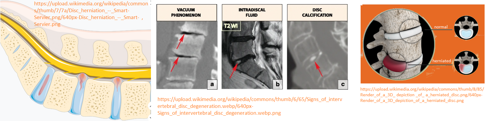
intervertebral disc components
Physio-Pedia has an excellent article of the intervertebral disc. The IVD consists of three major components: the central nucleus pulposus (NP), the peripheral annulus fibrosus (AF), and two vertebral endplates (VEPs) The NP is a completely bloodless tissue whereas the AF and VEP receive blood from by small branches from metaphysical arteries. Even this supply decreases as we age. All nutrients and oxygen that enter the NP and toxins that exit the NP, do so by diffusion. This may be the cornerstone of PEMF. Other thinking is that removal of dysfunctional IVD material might be another mechanism of PEMF in treating IVDD.
This youtube video shows materials with the same viscoelasticity as NP material being squished by a person’s thumb and forefinger. Texture of The Nucleus Pulposus – Dynamic Disc Design makes a model that helps practitioners with the textured biofidelity of the anatomical tissues of the intervertebral disc. This video lasts all of 15 seconds then explores herniating lumbar and cervical spine models as well as blood supply in other youtube videos by the same company Dynamic Disc Designs.. The Herneated Disc model even shows vascularization. The YouTube videos are a good place to start before moving on to the videos on the Dynamic Disc homepage. PEMF increases blood flow. The products of Dynamic Disc, if they represent reality, provide the practitioner something to think about. If the practitioner favors the Faraday Law of Induction mechanism of moving charged materials about, the mechanical properties of these models gives the practitioner something to ponder. ,

- The AF contains an inner and an outer portion. They differ primarily in their collagen composition. While both are primarily collagen, the outer annulus contains mostly type I collagen, while the inner has predominantly type II. A cartoon from this Physiopedia has been inlayed with a Wikimedia Commons scanning electron microscope image of type I collagen.
- The NP may contain type VI, IX, and collagens and proteoglycans. The proteoglycans include the larger aggrecan and versican that bind to hyaluronic acid, as well as several small leucine-rich proteoglycans. Aggrecan is largely responsible for retaining water within the NP. This structure also contains a low density of cells. While sparse, these cells produce the extracellular matrix (ECM) products (aggrecan, type II collagen, etc.) and maintain the integrity of the NP.
- The VEP permits diffusion and provides the main source of nutrition for the disc. It also contains a lot of collagen, of which there are thee types according to the Wikipedia cartilage page. Cartilage is composed of amounts of collagen and proteoglycan, proteins rich in saccharide based polymer modifications. The hyaline, endplate is also the last part of the disc to wear through during severe disc degeneration.
Fournier 2020 a reference from the Physio-pedia page on the IVD reviews blood supply to the three regions of the IVD.
copyright images of the intervertebral discs
Science Photo Library has some nice microscope images of the healthy intervertebral disc. Note the integrate nature of this structure. If one favors the Faraday Law of Induction simply getting charged particles to move, one can find it in these images.
a clinic using PEMF to treat IVDD in humans
A human clinic is using PEMF for IVDD. Dr Johnathan Vellinga of the Temecula Center for Integrative Medicine took the results from the Zeng 2022 study [1] and implemented in his clinic. Dr Vllinga’s post spells out why autophagy, “self eating”, is so important to remove damaged disc tissue.
PEMF vs Static magnet comparisons
After reviewing the background information, the reader probably has a visual of how crowded things are in our intervertebral discs. Faraday’s Law is that magnetic fields in motion get charged particles moving. Surely this aids in this “diffusion” that is part of the life of the IVD. Or does the magnetic field do something on its own? It would be nice o have a side by side comparison of a static and a PEMF by the same laboratory to address this question. Something close was found in two separate publications from different groups.
the introductions
PEMF [1]
IVD contains
- the central gel nucleus pulposus (NP) rich in aggrecan,
- collagen based annulus fibrosus (AF),
- the cartilage vertebral endplate (VEP) connecting vertebral bodies.
Changes in the extracellular matrix (ECM), inflammation, catabolism, and aging promote IVD degeneration. PEMF movement of ions on the cell membrane surface modulates proliferation, differentiation, and extracellular matrix (ECM) synthesis Previous work suggested sirtuin 1 (Sirt1) involvement. No summary cartoon was presented in this report. Sirt1 in autophagy came from a different publication. AMPK is a kinase activated when ADP/ATP ratios increase. Fig 6 from a previous publication shows the proposed signaling pathways of the effect of the Chinese medicine Yiqi Huoxue Recipe in IVDD.
static magnetic fields [2]
Secretion of proteoglycans within the extracellular matrix was claimed to be crucial for maintaining IVD homeostasis. Mitochondria dysfunction of nucleus pulposus cells (NPCs) was claimed to result from general wear and tear and inflammation. Sub optimal mitochondria means disruption of synthesis and metabolism of the extracellular matrix (ECM). This was more a technology development paper based on previous work of Marędziak 2015 that demonstrated that a 0.5T static magnetic field increases the synthesis and secretion of membrane-derived microvesicles (MVs) rich in VEGF and BMP-2 in equine adipose-derived stromal cells
Animal and cell models
| parameter | PEMF | static magnetic |
|---|---|---|
| animal | rat | rat |
| injury | puncture the tail disc from Co6-Co8 levels, including two IVDs (Co6-Co7 and Co7-Co8) | coccygeal IVD (Co 6/7) cutaneously punctured by a 21G needle at a depth of 5 mm, followed by rotation at 360° and holding for the 30s. |
| cultured cells | NP& mesenchymal stem cells | nucleus pulposus |
Devices
| parameter | PEMF | static magnetic |
|---|---|---|
| magnetic field | 0.3 mT | 40, 80 mT |
| frequency | 15 Hz details | NA |
| duration | 24, 48, 96hr cells | 72 hr |
The PEMF waveform comprised of a pulsed burst (burst width 5 ms; pulse width, 0.2 ms; pulse wait, 0.02 ms; burst wait, 60 ms; pulse rise and fall times: 0.3 µs, 2.0 µs), which was frequented at 15 Hz. This waveform is known to exert a therapeutic effect on nerve injury and osteoporosis in prior studies by the same group.
Results
| PEMF | static magnetic |
|---|---|
| Fig 2 In NPC PEMF decreased matrix metalloprotease mRNA and increased collagen mRNA. Less apoptotic cells. | Fig. 1 MSC co-culture delays NPC senescence. Markers were examined in IVDD rats too. ATP production was rescued in the cell culture model. |
| Fig 3 PEMF increases Sirt1 mRNA and mRNA of autophagy related proteins. | Fig. 2 MitoMVs, mitochondria containing micro vessels released by MSCs alleviated NPCs senescence and mitochondrial dysfunction. |
| Fig 4 Sirt1 inhibitors 3-MA inhibits the PEMF effect. | Fig. 3 SMF enhances mitoMVs secretion and further alleviates NPCs senescence. MitoMV increase as a function of field strength. |
| Fig 5 analyses of SIRT1 ⇑, Collagen II ⇑, MMP-3 ⤋, p62 ⇑, and LC3B ⇑ protein levels in NP cells ± Sirt1 inhibitor EX-527 inhibited the PEMF-mediated induction of ECM synthesis in NP cells, | Fig. 5 SMF enhances the interaction between Kif5b and Rab22a, and promotes the secretion of mitoMVs. Immuno precipitation, immunocolocalization, silencing RNA. See scheme 1 for a reminder of he role of the molecular motor Kif5b and Rab22 in vesicle transport on micro tubules and membrane fusion. |
| Fig 6 PEMF exposure improved rat IVD degeneration in vivo. | Fig. 6 departed from basic science and ventured into the world of creating a hydrogel production containing these mitochondria micro vesicles to treat IVDD |
| Fig 7. Rat IVD puncture causes lysosome LC3 and Sirt1 protein levels to fall. PEMF brings up the protein levels but not to control. | Fig 7 The mitochondria micro vesicle hydrogel in the in vivo rat IVDD model. |
In their discussion Zheng 2022 cited literature suggesting Faraday’s Law of Induction and the movement of charged particles without stating how this might impact Sirt1 activity and autophagy. Shi 2024 really did not care about how static magnetic fields might impact the secretion of microvessels containing mitochondria. One might think that the presence of a static magnetic field might impact the charges on a microtubule and hence the migration of kenesin carrying micro vessel cargo. Fun animation showing kinesin with cargo moving about a microtubule. This movement requires ATP! Static magnetic fields can orientation phospholipids covered in the membrane leaks fact checking post. There’s another explanation:
The Sirt1 Autophagy connection
Macroautophagy is catabolic membrane trafficking pathway that degrades cellular components through autophagosomes and lysosomes. Among many other things, it mediates the down regulation of nuleus mammalian SIRT1 protein during senescence and aging. [3] Importantly, the autophagy–lysosome pathway contributes to the loss of sirt1. [3] Kritschil 2023 demonstrated that autophagy is important for IVD health. [4]

Summary
The intervertebral disc is a very glycated, protein crowded space with limited blood flow that gets worse as we age. Surely proteins that contribute to the viscoelastic cushion need to be removed by macro autophagy. Zheng and coauthors demonstrated that PEMF activates this “self eating” authophagy that is so important to IVD health. Shi 2024 were looking for a way to use magnetic fields to rev up the export of mitochondria to help out stressed NP cells to have the energy to make more proteins. PEMF is reported to activate mitochondria ATP production. To get fancy, mitochondria complex II has been discussed as an antenna for PEMF on this site.. Nutrients and toxins are said to migrate in and out of the NP by diffusion. Does PEMF speed up this process? PEMF could be hypothesized to be a way of maintaining IVD health as we age..
A previous post PEMF and inflammation mentioned Tang 2017 a study a study that demonstrated that PEMF decreased IL‐1α induced IL‐6 Gene Transcription. In other words, one inflammatory cytokine tells the cell to make another inflammatory cytokine.
References
- Zheng Y, Mei L, Li S, Ma T, Xia B, Hao Y, Gao X, Wei B, Wei Y, Jing D, Luo Z, Huang J. Pulsed Electromagnetic Field Alleviates Intervertebral Disc Degeneration by Activating Sirt1-Autophagy Signaling Network. Front Bioeng Biotechnol. 2022 Mar 21;10:853872. . PMC free paper.
- Shi P, Gao H, Cheng Z, Zhao K, Chen Y, Chen X, Gan W, Zhang A, Yang C, Zhang Y. Static magnetic field-modulated mesenchymal stem cell-derived mitochondria-containing microvesicles for enhanced intervertebral disc degeneration therapy. J Nanobiotechnology. 2024 Jul 31;22(1):457. PMC free paper
- Xu C, Wang L, Fozouni P, Evjen G, Chandra V, Jiang J, Lu C, Nicastri M, Bretz C, Winkler JD, Amaravadi R, Garcia BA, Adams PD, Ott M, Tong W, Johansen T, Dou Z, Berger SL. SIRT1 is downregulated by autophagy in senescence and ageing. Nat Cell Biol. 2020 Oct;22(10):1170-1179. PMC free paper
- Kritschil R, Li V, Wang D, Dong Q, Silwal P, Finkel T, Lee J, Sowa G, Vo N. Impact of autophagy inhibition on intervertebral disc cells and extracellular matrix. JOR Spine. 2023 Sep 13;7(1):e1286. PMC free paper
Leave a Reply