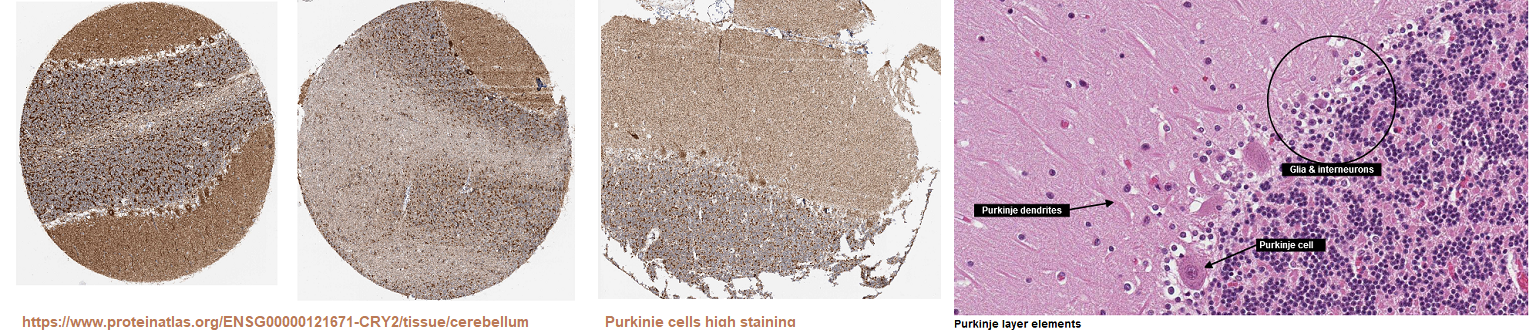
Most of this post is about work by Dr Rachel Sherrrard’s group at Sorbonne Université in Paris France and collaborators from around the world. The featured image is from Protein Atlas showing Cry2 staining in the cerebellum. The very obviously striated staining for Cry2 in the cerebellum and long with biochemical proof of Cry2 involvement in the PEMF and static magnetic field response make these collaborations truly insightful. Now that we have more proof of a PEMF target we need to start taking it more seriously.
- PEMF can cause cryptochromes to generate H2O2 in cultured cells. Low level magnetic fields increase the transcription of three reactive oxygen species sensitive genes.
- Mice lacking Cry1 and Cry2 did not respond to trans cranial low frequency magnetic fields. The target is healing of severed olivo-cerebellar connections. Purkinje cells, rich in Cry2, establish connections to climbing fibers.
- A 2023 Sherrard Group review presented multiple mechanisms of magnetic fields for neuro circuit repair. Links to insightful cartoon illustrations are provided.
References
Previous work by Sherrard, Ahmad, and others [1, 2]
Dr. Margaret Ahmad, a plant scientist, was a key force in these two publications. The Ahad group’s prowess in cryptochrome biochemistry in plants and animals is documented in “Seeing” the Electromagnetic Spectrum.
Cry1/2 is needed for PEMF production of H2O2 [1]
Two common mammalian cell lines, HEK 293 and MEF, were exposed to a frequency of 10 Hz and 2 mT. 1 cm below he culture dish. [1] PEMF was found to induce generation of intracellular H2O2 in a cryptochrome dependent manner. [1]
- Fig 1 fly behavioral and cellular response to PEMF. Panel A shows distribution of wild-type Canton S larvae following 96-hour exposure in blue light to activate cryptochromes and PEMF (10 Hz) at the lower left corner. The larvae at at the upper right corner. Flies expressing human Cry1 do this too. Cry deletion mutants do not.
- Fig 2 PEMF response of HEK293 cell cultures. The strategy was to take a common cell line and knock down the expression f both Cry1 and Cry2. Instead of no PEMF, PEMF in apposing directions so as to cancel out magnetic fields was an added control. PEMF and Cry are needed for the production of H2O2 and the increase in number of cells.
- Fig 3 This figure contains some very nice images of a fluorescent dye that detects H2O2 in two common cell lines exposed to PEMF. parallel the imaging data of these HEK293 cells, which showed enhanced localization of ROS to the nuclear, Golgi, and ER compartments.
Induction of charged particle flow or radical pair mechanism? [2]
In a follow up study HEK293 cells were exposed to low level fields: control (40 μT), 500 μT or 2000 μT static fields for either 10 minutes or 3h, [2] Static fields had little effect on gene expression. [2] The control (40 μT) appears to be the magnetic field of the incubator. Cryptochromes seem to be a likely antenna for PEMF. They are also the way plants and animals sense the magnetic field of the earth. Is PEMF simply canceling out the latter?
- 2 mT was the peak intensity of magnetic field output by the PEMF device used in the previous study showing induction of ROS. [1]
- 500 μT induces physiological effects and modulation of cryptochrome function in other models. This is 10x the earth’s magnetic field according to the authors.
- low-level field LLF (less than 200 nT intensity) they chose 40 μT the magnetic field inside the inside the incubator. Cells are effectively deprived of the earth’s magnetic field by placing within two concentric μ-metal cylinders with walls 0.2 mm thick,. . Local magnetic fields at the center of the μ-metal cylinder are lower than 200 nT.
Fig 1 Effect of low level field (LLF) on HEK293 gene expression. [2]
- KIAA1211 is involved in epithelial cell integrity by acting on the dynamics of the actin cyto skeleton by regulating actin polymerization and inhibiting the interaction of actin-capping proteins. A 10 minute exposure increased the level of these transcripts 2.5x. The implication if more filamentous actin.
- RPS16P5 is a smaller component of the small ribosomal subunit. Ribosomes translate messenger RNA into proteins. Exposure for 10 minutes and three hours doubled the transcripts for this protein implying that the exposed cells might be better at translating mRNA into proteins.
- TAS2R19 is the bitter taste receptor also known as gustducin. The implication in increases of this transcript is unknown.
Discussion of the discussion
This exciting publication presented many difficult concepts. The discussion has been reduced to bullet points with additional comments.
- The initial expectation was that more magnetic field exposure would create more effect. The concept of just a little ROS stimulating oxidative stress gene expression was covered in the ozone therapy post. ROS–regulated gene expression was identical at all three tested static magnetic field conditions—40 μT, 0.5 mT and 2 mT. Only the absence of the magnetic field stimulated a significant increase in ROS-regulated gene expression.
- ROS synthesis has an optimal biological minimum at the intensity of the Earth’s geomagnetic field, otherwise higher magnetic field strengths would have biological consequences.
- The magnetic component of PEMF sessions cover a range of directional vectors and intensities (rising and falling) at rates determined by the PEMF pulse frequency and signal shape. But wouldn’t the receptor of such magnetic fields also be reorientating via Brownian motion of the cellular milieu ?
- It must also be considered that magnetic fields are additive and therefore can cancel each other out when coming from different directions at comparable intensities.
- Over a prolonged PEMF exposure period, all of the susceptible receptor molecules throughout the sample would be intermittently exposed to the LLF.
- Transient static LLFs formed in the course of PEMF stimulation could thereby significantly impact on rates of ROS formation in cell culture.
- For instance, if each flavoprotein receptor molecule received a single transient LLF stimulus at least once during the (10 min or 3 hr)… This is getting way too complicated. We’ve got think about other factors involving protein thiol groups that might be sensitive to the H2O2 being generated.
- Radical Pair mechanism is that quantum physical effects of the magnetic field on electron spin chemistry are predicted to operate in a narrow range and reach a plateau beyond which further increase in magnetic field intensity could have no further effect on product yields, i.e. biological response at Earth magnetic field (40 μT) are similar to 0.5 and 2 mT.
- The possibility of induced electric fields by PEMF was acknowledged.
The next publication explored PEMF frequencies approximating brain wave frequencies for the purpose of repairing sevred circuits in a cryptochrome knock out mouse model.
Cry2 is an antenna for repetitive trans-cranial magnetic stimulation evoking brain repair [3]
According to these authors commonly used non invasive brain stimulation protocols use electromagnetic stimulation in one of two forms:
- strong electromagnetic pulses 0.5 to 2 Tesla. The idea is to depolarize neurons
- low-/pulsed-field magnetic stimulation microtesla (μT) to millitesla (mT) delivered to the whole brain that modulate release of neurotrophic factor (BDNF) and other growth factors. Increased intracellular Ca2+ is part of the process.
Most of the figures in this publication are going to be left to the reader to understand. Many of the tactics seem to mimic clinically used techniques.
protocol briefs
Dufor 2019 used a double knockout Cry1−/−Cry2−/− mouse model to test the hypothesis that cryptochrome are the antenna. The olivocerebellar axonal connection was transected. Mice were allowed to recover before dividing into treatment groups.
Fig. 1 LI-rTMS induces transcommissural climbing fiber re-innervation in injured adult mice.
This figure explores the concept of climbing fibers and VGLUT2 …The vesicular glutamate transporter is actually a uni porter for many ions and small molecules important for the functioning of neurons. Note that the field intensities are larger than some of those tested in cell culture studies. [2] The online version of this image does not magnify well. Recommended saving as a PDF file and enlarging the image that way. You, the reader, decide if BGLUT2 staining compares with Cry2 staining in the human cerebellum in the featured image.
Wikipedia authors have compiled a good page on Climbing Fibers, are are able to undergo repair modifications in response to injuries by forming new branches to innervate surrounding Purkinje cells. Climbing fibers are connected to the inferior olivary nucleus (ION) that coordinates signals from the spinal cord to the cerebellum
Fig. 2 LI-rMS pattern regulates climbing fiber re-innervation ex vivo.
The Wikipedia page on Purkinje cells mentions the Purkinje cell protein 4 that facilitates the calcium calmodulin association and dissociation. Simple spikes (7-150 Hz) and complex spikes (1-3 Hz) were also mentioned in this Wikipedia page.
Does the extremely low frequency PEMF mimic normal rhythms of these synaptic connections? Note we are back to Pooam 2020 [2] in the previous section. The field intensities are larger than the very weak ones shown to alter three selected gene transcripts.
Fig 3 LI-rMS intensity has a lower limit below which re innervation fails.
Figure 3 concluded that repetitive magnetic stimulation to the target cerebellum must be above a 4.5 mT baseline intensity to induce olivo-cerebellar re-innervation.
Fig 4 LI-rMS modulates gene expression in denervated hemi-cerebellar plates
In attempts to take a biochemical approach to a very neuro anatomy publication, panel 4D offers some insight: percentage of calbindin- or parvalbumin-positive cells that are double-labeled with c-fos. Note that calbindin and parvalbumin are calcium handling genes rather than oxidative stress related genes.
- c-fos is a transcription factor that forms hetero-dimers with c-Jun, aka the AP1 complex. Wikipedia authors discuss the use of c-fos as a measure of neuronal activity.
- Calbindin is a vitamin D dependent calcium binding proteins with EF hands like calmodulin. Wikipedia authors report abundance in the cerebellum.
- Parvalbumin is also a small calcium binding protein with EF hands like calmodulin.
Fig 5 Cryptochromes are required for LI-rMS–induced post lesion repair.
The take home message seems to be that the interaction of an intact brain, or ex-planted parts of an entire brain, are very complicated. The presence of Ca2+ binding proteins, perhaps only as markers, reinforces the notion that PEMF, reactive oxygen species, and changes in intra cellular ca2+ are doing something. Apologies to the authors for not doing the neuro anatomy justice.
Cry2 and other PEMF hypotheses [4]
Fig 1 is a cartoon showing the trans cranial setup. Panel C shows the inside negative membrane along the length of the axon. There is a small region that has depolarized with an inside positive membrane potential. The following image is a non copy righted view of these regions of depolarization or hyper polarization that PEMF might be affecting.

Wikipedia authors have an excellent page on Saltatory Conductance that makes use of these Nodes of Ranvier. The intriguing aspect of this work is that PEMF might aide this process.
Fig 2 shows a “figure 8” coil system that delivers high field intensity in some anterior brain “hot spots.” In general, anterior regains of the brain receive less of a field. It is interesting to note that the field strength is presented in units of V/m instead of Tesla’s.
Fig 3 shows experimental devices to deliver PEMF to mouse brains. In this case field strength is presented in units of mT.
Fig 4. Illustrates the concept of pulse patterns, frequencies, and shapes for the purpose of targeting specific regions within the brain.
Fig 5 gives an overview of the favorite antenna for PEMF: cryptochrome. The Ca2+ activated by reactive oxygen species generated by activated FAD-bound Cry is not specified.
Fig 6 offers an alternative hypothesis as to how PEMF might enhance new gene transcription. PEMF acts on L-type voltage gated calcium channels. Glutamate acts on NMDA receptors. Both increase intracellular Ca2+ that activates the cyclic AMP response element (CRE) binding protein Creb. Downstream of the CRE is the gene for the transcription factor c-Fos. c-Fos activates the transcription of late response genes for long term plasticity.
What does the magnetic field do beyond moving charges? [5]
The official title of the abstract, “What does the magnetic field do? Neural changes beyond induction of an electric field.” is a good for those who favor Faraday’s Law of Induction to explain very low frequency PEMF. These are some bullet points from a Sherrard abstract found on Research Gate.
- We need animal models because structural changes in the human brain are difficult to measure. These changes include the neurons and their networks.
- This group is also using 3D organotypic culture and in primary cultures, over a range of stimulation parameters.
- The structural changes in the neurons themselves and their connections depend on the stimulation-pattern.
- Cryptochromes are the favored cellular magneto-receptor.
- Reactive oxygen species and increased intra cellular calcium are the favored second messengers for the expression of neuroplasticity-related genes.
- The Sherrard group hopes to personalize treatment strategies for brain repair.
References
- Sherrard RM, Morellini N, Jourdan N, El-Esawi M, Arthaut LD, Niessner C, Rouyer F, Klarsfeld A, Doulazmi M, Witczak J, d’Harlingue A, Mariani J, Mclure I, Martino CF, Ahmad M. Low-intensity electromagnetic fields induce human cryptochrome to modulate intracellular reactive oxygen species. PLoS Biol. 2018 Oct 2;16(10):e2006229. PMC free paper
- Pooam M, Jourdan N, El Esawi M, Sherrard RM, Ahmad M. HEK293 cell response to static magnetic fields via the radical pair mechanism may explain therapeutic effects of pulsed electromagnetic fields. PLoS One. 2020 Dec 3;15(12):e0243038. PMC free paper
- Dufor T, Grehl S, Tang AD, Doulazmi M, Traoré M, Debray N, Dubacq C, Deng ZD, Mariani J, Lohof AM, Sherrard RM. Neural circuit repair by low-intensity magnetic stimulation requires cellular magnetoreceptors and specific stimulation patterns. Sci Adv. 2019 Oct 30;5(10):eaav9847. PMC free article
- Dufor T, Lohof AM, Sherrard RM. Magnetic Stimulation as a Therapeutic Approach for Brain Modulation and Repair: Underlying Molecular and Cellular Mechanisms. Int J Mol Sci. 2023 Nov 17;24(22):16456. PMC free articles
- Sherrard, Rachel & Fauquier, Aurélien & Escriou, Guillaume & Dufor, Tom & Doulazmi, Mohamed & Lohof, Ann. (2023). What does the magnetic field do? Neural changes beyond induction of an electric field. Brain Stimulation. 16. 161. 10.1016/j.brs.2023.01.141. link to abstract
Leave a Reply