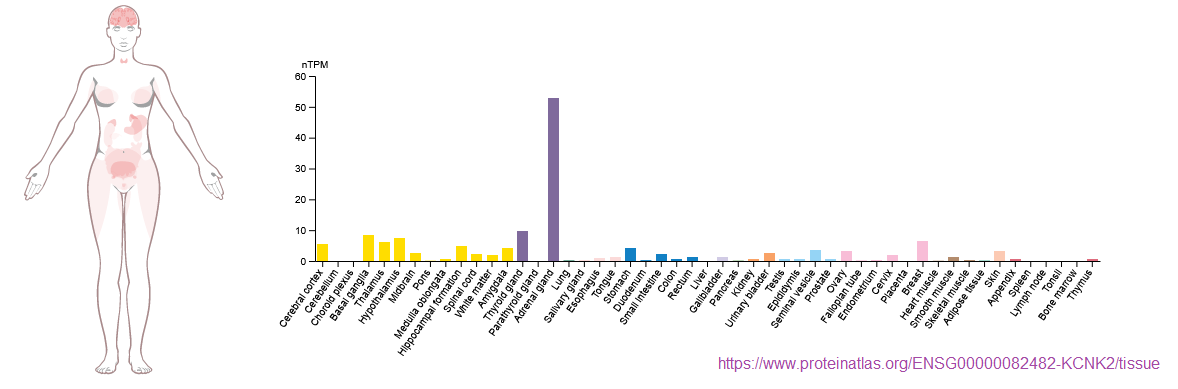
The featured image comes from ProteinAtlas that has something interesting to say about the organs in which TREK1 is expressed. It is not such a brain protein. According to UniProt.og ” TREK1 is an ion channel that contributes to passive transmembrane potassium transport. Reversibly converts between a voltage-insensitive potassium leak channel and a voltage-dependent outward rectifying potassium channel in a phosphorylation-dependent manner. In astrocytes, forms mostly heterodimeric potassium channels with KCNK1, with only a minor proportion of functional channels containing homodimeric KCNK2. In astrocytes, the heterodimer formed by KCNK1 and KCNK2 is required for rapid glutamate release in response to activation of G-protein coupled receptors, such as F2R and CNR1 (By similarity).”
Magnetic activation (1 Hz 25 mT) of TREK1 (via coupled nanoparticles) triggers stress signalling and regulates neuronal branching in SH-SY5Y cells. There might have been some influence of the PEMF alone
A very low frequency PEMF and TREK1
Rotherham M, Moradi Y, Nahar T, Mosses D, Telling N, El Haj AJ. Magnetic activation of TREK1 triggers stress signalling and regulates neuronal branching in SH-SY5Y cells. Front Med Technol. 2022 Dec 5;4:981421. PMC free paper
This study was using magnetic nanoparticles with antibodies against TREK1 to induce mechanical stress on the TREK1 K+ channel. Permanent ≥25 mT permanent NdFeB magnets where set to oscillate at 1 Hz for one our sessions. SH-SY5Y neuron like cells cells were grown to confluence. This very low frequency high field PEMF tended to decrease endothelial nitric oxide synthase expression. There was weak suggestion of PEMF alone increasing neurite outgrowth. Whether or not the voltage gated K+ channel TREK1 had anything to do with it is unknown because TREK1 expression was never “knocked out.” Could 25 mT in and of itself have caused mechanical stimulation of TREK1?
Bandulik S, Tauber P, Lalli E, Barhanin J, Warth R. Two-pore domain potassium channels in the adrenal cortex. Pflugers Arch. 2015 May;467(5):1027-42. PMC free paper
What are the roles of TREK1 and TASK potassium channels in the adrenal cortex? This seems to be the main site of expression. The Bandulik review is more focused on the TASK2 potassium channel in glomerulosa cells of the adrenal gland. These cells produce sodium conserving aldosterone in responsible to angiotenin II. The role of this renin-angiotensin -aldosterone =system is to preserve fluid volume. Anti-hypertension drugs target this axis. The fasiculata cells produce cortisol in response to angiotensin II. angII has the effect of inhibiting TREK1 with the effect of preventing this potassium from hyperpolarizing the cell. This depolarization opens voltage gated calcium channels. According to this model, calcium signaling ensues and stress hormone cortisol is produced.
See Fig 1
Do changes in fluid volume constitute a mechanical stress that can activate TREK1? One would speculate that such a scenario would prevent excess cortisol production in the face of activation of the RAAS to maintain fluid volume of our bodies. Could low frequency PEMF active adrenal gland TREK1 so as to decrease cortisol production?
Leave a Reply