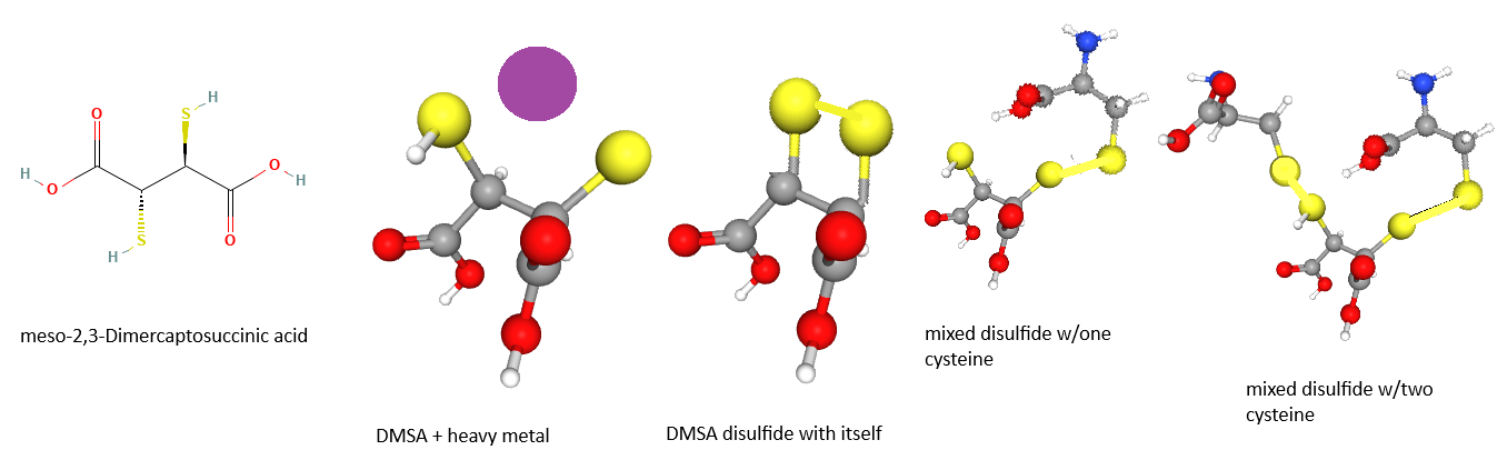
A colleague was interested in DMSA for treating patients with suspected heavy metal poisoning. There are a few clinics in the world that use DMSA for heavy metal detox purposes. In earlier decades dimercaptosuccinic acid (DMSA/succimer) was a wonder drug for treating lead and mercury poisoning. DMSA has two carboxyl groups and two thiols that are used for the chelation of “heavy metals.” DMSA seems to have fallen out of favor, despite our ever increasing toxin world. This post takes a brief look at the use of technium-DMSA as an imaging agent and gives a short bullet point review of the literature of heavy metal detox studies.
How DMSA is used to test renal function
DMSA can chelate a short half life radioactive element called technium. It has one free thiol left that it uses to form a disulfide bond with a thiol of just one of many plasma proteins that get filtered through the glomerulus. In a properly functioning kidney, filtered proteins are reabsorbed by megalin and cubulin.
“We describe a simple method of pentavalent technetium-99m dimercaptosuccinic acid [99mTc(V)-DMSA] preparation for the imaging of medullary carcinoma of the thyroid using commercially available kits. 99mTc(V)-DMSA is available at high pH (approximately 7.5) by adding NaHCO3 solution in the presence of a small amount of reducing agent (SnCl2). On the other hand, trivalent 99mTc-DMSA [99mTc(III)-DMSA] can be obtained at low pH (below 3) in the presence of an excess amount of reducing agent. In the clinical evaluation of a patient with a medullary carcinoma of the thyroid, only 99mTc(V)-DMSA revealed an area of intense accumulation.” (Hirano 1994) What this says about DMSA used for detox is that the binding may depend on the pH and the charge of the toxic element we wish to eliminate.

This PubChem style image was adapted from Grierosu 2025 Fig 11. Technium is cyan, sulfur is yellow, oxygen is red, carbo gray, and hydrogen white.
Note that there is only one free thiol (sulfur) not bound to technium that is available to bind to serum proteins. If one is to use DMSA to chelate heavy metals, it makes sense as to why one mixed disulfide with cyteine is a good thing such that heavy metal chelated DMSA doesn’t reenter the circulation.
de Lange 1989 made the case for glomerular filtration and peri-tubular reabsorption of Technium-99m-dimercaptosuccinic acid. In 13 individuals with normal renal function, the renal clearance of [99mTc]DMSA was measured by adding [99mTc] DMSA to the infusion fluid during the continuous infusion of [II31]hippurate and [I125]iothalamate for the measurement of effective renal plasma flow(ERPF) and glomerular filtration. The introduction of this old paper has some excellent points
- Technetium-99m DMSA flux through the proximal tubule by glomerular filtration, reabsorption, or uptake from the peritubular capillaries. Secretion was not mentioned.
- DMSA is largely bound to serum proteins which was measured by precipitation of aid serum protein with trichloroacetic acid or ultra-filtaration.
- Low renal uptake of [99mTc]DMSA was found in patients with defects in protein renal reabsorption .
Megalin/Cubulin: 99mTc-DMSA transporter Weyer 2013
By 2013 Dimercaptosuccinic acid (DMSA) labeled with 99mTc was a major renal cortical imaging agent used in the diagnosis of renal parenchymal disorders. Megalin and Cubulin are two transmembrane proteins that capture proteins in the lumen of the renal tublules and stimulate endocytosis of immunogobins, albumin, and one would think other stuff. Click here to view a Research Gate cartoon that is probably copyrighted. These authors used knockout mice to confirm the participation of these two transporters in reabsorption of the 99mTc amicroglobulin complex. The analysis of urine samples collected from megalin/cubilin-deficient mice by SDS-PAGE and autoradiography showed that the 99mTc radioactivity was associated with a single band of approximately 27 kDa. To identify the protein associated with the tracer, the collected urine samples were subjected to SDS-PAGE, and proteins were visualized by silver staining. The sample representing megalin/cubilin-deficient mice presents unique staining at the position corresponding to 27 kDa (Fig. 4B). This band was excised, subjected to in-gel digestion using trypsin, and subsequently identified as the plasma protein α1-microglobulin by MALDI MS
Fig 4B Urinary excretion of protein-bound 99mTc-DMSA by megalin/cubilin-deficient mice.
(A) SDS-PAGE separation and autoradiography of urine from megalin/cubilin-deficient mice and controls injected with 99mTc-DMSA. A single, labeled band of approximately 27 kDa is observed in urine collected from megalin/cubilin-deficient mice only. Note that only one protein is radioactive.
(B) Urine profile analysis by SDS-PAGE and silver staining of urine from megalin/cubilin-deficient mice and controls. A distinct protein band of 27 kDa was identified in urine from megalin/cubilin-deficient mice (*). This band was excised and protein identified by MALDI MS as α1-microglobulin. The authors used a protein stain to visualize all of the filtered proteins. Note that most of these proteins are not associated with any radioactivity. This means that DMSA may form weak mixed disulfides with other serum proteins, these interactions are likely transient.
(C) Immunoblotting identifying α1-microglobulin as an approximate 27-kDa band in urine from megalin/cubilin-deficient mice but not from controls. α1-microglobulin is a heme iron containing protein that has (1) Antioxidant and tissue repair protein with reductase, heme-binding and radical-scavenging activities (2) Reduces extracellular methemoglobin, a Fe3+ (ferric) form of hemoglobin that cannot bind oxygen, back to the Fe2+ (3) Catalyzes the reduction of carbonyl groups on oxidized collagen fibers and preserves cellular and extracellular matrix ultrastructures, and (4) acts as a protease inhibitor.
Fig 6 shows a cartoon of how the authors think things happen. The are not sure of the fate of megalin/cubilin endocytosed α1-microglobulin. It does not matter because it is essentially the only serum protein with stable binding.

A quote from PDB “We describe the 2.3 Å (1 Å=0.1 nm) X-ray structure of α1m (α1-microglobulin), an abundant protein in human blood plasma….Loop #1 harbours the residue Cys34 which is responsible for covalent cross-linking with plasma IgA. A single disulfide bond between Cys72 and Cys169 connects the C-terminal segment to the β-barrel, as in many other lipocalins. The exposed imidazole side chains of His122 and His123 in loop #4 give rise to a double Ni2+-binding site together with a crystallographic neighbour. In contrast with these, α1m is initially expressed as a bifunctional fusion protein with the protease inhibitor bikunin. Neither the electron density nor ESI-MS (electrospray ionization MS) provide evidence for a chromophore bound to the recombinant α1m, also known as ‘yellow/brown lipocalin’ ands accommodated in the hydrophobic pocket. A structural feature similar to the well-known Cys-Pro haem-binding motif indicates the presence of a haem-binding site within the loop region of α1m, which explains previous biochemical findings and supports a physiological role in haem scavenging, as well as redox-mediated detoxification.”

Here we have the same crystal structure viewed by the software of rcsb.org that allows us to view water molecules surrounding Ni that is liganded to some histidines. Many proteins in general may have amino acids in the right place to bind heavy metals.
Just a point to come back to later: Perhaps heavy metals bound non-specifically to serum proteins like cadmium is bound to this crystal structure of microglobulin is a route of detoxification.
A few snap shots of DMSA used for heavy metal detox
There is absolutely no way this post is going to review several decades of DMSA research. Sadly, much of this research was performed before the days of high powered mass spectrometers.
- Kobayashi 2021 concluded that the basolateral organic anion transporter 1 (OAT3) was responsible for transport of [99mTc]DMSA. [99mTc]DMSA accumulates in renal proximal tubular epithelial cells from blood via OAT3 on thebasolateral side, and then a small volume of [99mTc]DMSA will be excreted in urine via MRP2 My question is where mixed disulfide formation with mciroglobulin occurs and what percentage is excreted before protein disulfides can form.
- Burckhardt 2002 acknowledged the role of the 3Na+ dicaboxylate transporter 1 (NaDC1) for active transport of Krebs cycle intermediates such as succinate, a-ketoglutarate, and citrate. NaDC1 is found in the luminal (NaDC-1)and basolateral plasma membranes (NaDC-3) of proximal tubule cells. This was an electrophysiology study using flounder NaDC expressed in frog oocytes. fNaDC-3 transported 2,3-dimethylsuccinate and so on. A critical role of theposition of the methyl- or sulfhydryl-group in voltage-sensitive affinity was determined. The chelates formed by HgCl2 and DMSA or DMPS and by Pb(NO3)2 and DMSA, however, were not translocated by fNaDC-3. NaDC-3 was concluded to be an essential component in the delivery of uncomplexed antidotes for renal heavy metal detoxification but not the complex themselves. So does the DMSA-Me complex in the srum just get filtered and not reabsorbed?
- By 14 hr after DMSA administration (10 mg/kg), only 2.5% of the administered DMSA is excreted in the urine as unaltered DMSA and 18.1% of the dose is found in the urine as altered forms of DMSA. Most altered DMSA in the urine is in the form of a mixed disulfide. It consists of DMSA in disulfide linkages with two molecules of L-cysteine. One molecule of cysteine is attached to each of the sulfur atoms of DMSA. The remaining 10% of the altered DMSA was in the form of cyclic disulfides of DMSA. So far, the mixed disulfide has been found in human but not in rabbit, mouse, or rat urine.(Aposhian1990) See the featured image. My question is where this mixed disulfide forms? Is it in the kidney or in the serum? I would have predicted a mixed disulfide with glutathione.
- A typical course of oral DMSA consists of 30 mg/kg body weight/day, for 5 days,often followed by 20 mg/kg/day for 14–21 days. after oral administration to healthy adult volunteers, “total” DMSA is distributed in the body’s extracellular fluid and in blood. In the latter, total DMSA is almost entirely distributed in the plasma and does not penetrate erythrocytes.[10,12] Very little “unaltered” DMSA is found in plasma. Total DMSA consists of unaltered DMSA plus oxidized (disulfide) DMSA; altered DMSA is the difference 92% of the DMSA is bound to albumin. (Asiedu 1995)
- Two monoesters of meso-2,3-dimercaptosuccinic acid (DMSA), monoisoamyl meso-2,3-dimercaptosuccinate (Mi-ADMS) and mono-n-hexyl meso-2,3-dimercaptosuccinate (Mn-HDMS) were compared to DMSA in their efficiency to mobilize 203Hg in mercury-laden suckling rats. Seven-day-old pups were given 203Hg (18.5 kBq) with a dose of 0.5 mg Hg/kg/day as HgCl2 for five days. Seven days after the beginning of Hg loading a ten-day oral treatment with DMSA, Mi-ADMS, or Mn-HDMS was administered at a dose of 0.25 mmol/kg/day. At the end of experiment, radioactivity was measured in the whole body, liver, both kidneys, and brain. (Kostial 1995)
- DMSA is an effective lead chelator that primarily chelates renal lead. It is generally well tolerated but may occasionally cause clinically important adverse effects. DMSA may now be considered as an alternative to sodium calcium edetate, particularly when an oral antidote is preferable. (Bradbury 2009)
What very little we know
The colleague who was originally interested in use of DMSA for patients with long term and unknow heavy metal accumulation read all they could on the use of DMSA for heavy metal toxicity and probably became the world’s leading expert on this topic. They informed me that this model I came up with to reconcile the uses of DMSA was wrong. The same colleague decided to go with a novel twist on a different chelation protocol because DMSA “goes to the bone.” I’m not going down this rabbit hole that the colleague has decided to move on from. Here is my literature based model, that I agree with my colleague, is probably wrong.
- Glomerulus filtered proteins are reabsorbed by the megalin/cubulin complex. This could include 99mTc-DMSA covalently bound to microglobulin (Weyer 2013). I’m informed that even though little albumin is filtered, DMSA can bind to albumin with or without a heavy metal or not. Or heavy metals can just bind to serum proteins without DMSA. Then they get digested by lysosme upon endocytosis.
- We know that DMSA in the urine in conjuated to 1-2 cysteines. (Aposhian1990) What is with this? We have no reason to doubt this whatsoever, but glutathione conjugates would have been the predicted result. The featured image makes a case for conjugate being a better chelator.
- [99mTc]DMSA is absorbed by the blood via OAT3 and then excreted via MRP2 (Koybayashi 2023). I’m told this is not the same complex as DMSA conjugated to a divalent heavy metal rather than a pentavalent heavy metal. Even Hirano 1994 informs us we get a different Tc (V) complex with DMSA depending on the pH and the charges on carboxyls and cysteines.
- Then Burckhardt 2002 informs us that unconjugated DMSA gets into proximal tubule cells via NaDC1. Naturally heavy metal conjugation would totally screw up the charge gradient that powers this transport.
- Now how does unconjugated DMSA get into the lysosomes once they are fused with the endosome? Certainly this would be a good source of free cysteines and whatever heavy metals might be stuck nonspecifically to filtered serum proteins. Fine. Then how to they get out of the lysosome to hopefully be transported out by mrp2?

This model has got to be wrong! Perhaps the decision to go with another chelator was based on not knowing the right model. Logistic of use of DMSA is certainly another factor. Unlike my colleague, I’ve not skimmed though most, if not all, DMSA studies every published. Knowing what I do about the tendency of protein thiols to form mixed disulfides, I see the lack of the sophisticated mass spectrometry when DMSA detox research was not as a draw back and a caution.
References
- Aposhian HV, Aposhian MM. meso-2,3-Dimercaptosuccinic acid: chemical, pharmacological and toxicological properties of an orally effective metal chelating agent. Annu Rev Pharmacol Toxicol. 1990;30:279-306. PubMed
- Asiedu P, Moulton T, Blum CB, Roldan E, Lolacono NJ, Graziano JH. Metabolism of meso-2,3-dimercaptosuccinic acid in lead-poisoned children and normal adults. Environ Health Perspect. 1995 Jul-Aug;103(7-8):734-9. PMC free article
- Bradberry S, Vale A. Dimercaptosuccinic acid (succimer; DMSA) in inorganic lead poisoning. Clin Toxicol (Phila). 2009 Aug;47(7):617-31. free paper
- Burckhardt BC, Drinkuth B, Menzel C, König A, Steffgen J, Wright SH, Burckhardt G. The renal Na(+)-dependent dicarboxylate transporter, NaDC-3, translocates dimethyl- and disulfhydryl-compounds and contributes to renal heavy metal detoxification. J Am Soc Nephrol. 2002 Nov;13(11):2628-38. doi: 10.1097/01.asn.0000033463.58641.f9. PMID: 12397032.
- Grierosu IC, Starcea IM, Jalloul W, Mocanu MA, Bogos RA, Lazaruc TI, Beldie MA, Tibu R, Ionescu TM, Stolniceanu CR, Acsinte BC, Stefanescu C, Saviuc A, Ghizdovat V. Particularities in the “Oldie but Goldie” Tc-99m DMSA Renography: A Retrospective Reference Centre Overview of 931 Children. Diagnostics (Basel). 2025 Apr 17;15(8):1025. PMC free paper
- Hirano T, Tomiyoshi K, Zhang YJ, Ishida T, Inoue T, Endo K. Preparation and clinical evaluation of technetium-99m dimercaptosuccinic acid for tumour scintigraphy. Eur J Nucl Med. 1994 Jan;21(1):82-5. PubMed
- Kobayashi M, Mizutani A, Okamoto T, Muranaka Y, Nishi K, Nishii R, Shikano N, Nakanishi T, Tamai I, Kleinerman ES, Kawai K. Assessment of drug transporters involved in the urinary secretion of [99mTc]dimercaptosuccinic acid. Nucl Med Biol. 2021 Mar-Apr;94-95:92-97. Sci-Hub free paper
- Kostial K, Blanusa M, Piasek M, Jones MM, Singh PK. Prolonged oral treatment with two monoesters of meso-2,3-dimercaptosuccinic acid for depleting inorganic mercury retention in suckling rats. Pharmacol Toxicol. 1995 Sep;77(3):216-8. PubMed
- de Lange MJ, Piers DA, Kosterink JG, van Luijk WH, Meijer S, de Zeeuw D, van der Hem GK. Renal handling of technetium-99m DMSA: evidence for glomerular filtration and peritubular uptake. J Nucl Med. 1989 Jul;30(7):1219-23. PMC free article
- van Eijkeren JC, Olie JD, Bradberry SM, Vale JA, de Vries I, Meulenbelt J, Hunault CC. Modelling dimercaptosuccinic acid (DMSA) plasma kinetics in humans. Clin Toxicol (Phila). 2016 Nov;54(9):833-839.
- Weyer K, Nielsen R, Petersen SV, Christensen EI, Rehling M, Birn H. Renal uptake of 99mTc-dimercaptosuccinic acid is dependent on normal proximal tubule receptor-mediated endocytosis. J Nucl Med. 2013 Jan;54(1):159-65. free article
Leave a Reply