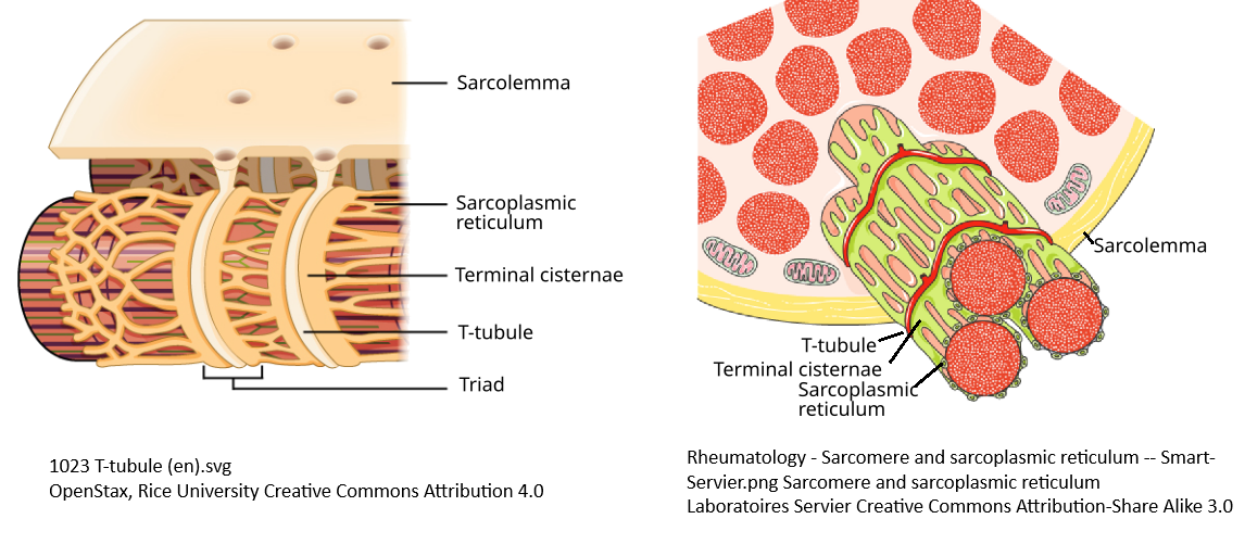
Many of the high power PEMF device practitioners see muscle twitching, tiny little contractions, as evidence that PEMF is giving the tissue a “cellular workout.” Many claim that voltage gated calcium channels are opened by PEMF. Some practitioners are completely and totally without a clue as to whole complicated things are. The featured image contains the views of two artists of the triad of membranes that. The voltage gated Ca2+ Cav1.1 resides in the t-tubules. This post introduces some other voltage gated participants in excitation contraction coupling. Real life is far more complicated than this post too! Geometry matters too! You have no clue how the orientation of your coils impacts these three dimensional membrane structures. The goal is cautionary when it comes to use of high power PEMF devices that are strong enough to cause muscle twitching.
Overview
These are the topics to be covered in this post

- The basics of skeletal muscle contraction
- Overview of ion transporters
- The Na+ Pump, aka the Na/K-ATPase, indirectly PEMF sensitive
- Ca2+ ATPases , indirectly PEMF sensitive
- CaV1.1 and the ryanodine receptor, gating is very complicated
- Voltage gated K+ and Na+ channels, some PEMF sensitive
- TRPC1 & Cry2, PEMF sensitive
- ClC1 the chloride channel, does have a voltage gate
- The Na+/Ca2+ exchanger, PEMF sensitive
- Tying transporters together
The basics of skeletal muscle contraction
Which steps can be influenced by PEMF? We’ll conclude that the coupling between DHP/Cav1.1 to the ryanodine receptor in the sarcoplasmic reticulum is grossly over simplified in this cartoon.

- The motor neuron releases a neurotransmitter
- An action potential wave travels into the t-tubule
- The voltage gated L-type CaV1.1, aka the dihydropyridine receptor, opens to let Ca2+ into the cell.
- CaV1.1 is also mechanically coupled to the ryanodine receptor which releases Ca2+ sarcoplasmic reticulum storage.
- Ca2+ binds to troponin C of the troponin complex.
- Activated troponin grabs tropomyosin out of myosin’s way.
- ATP hydrolysis by myosin heads generates force.
Ion channels and transporters discussed in this post
Imagine a three dimensional version of this cartoon. The t-tubule is a cylindrical hole in the surface of the muscle fiber. The sarcoplasmic reticulum, that stores Ca2+ is also three dimensional with CERCA ATP driven Ca2+ pumps over the surface. Cav1.1 is a voltage gated Ca2+ channel.


Perhaps the reality is that charged particles move in a swirling perpendicular motion relative to the magnetic field. If we are dealing with three dimensional structures, what does this mean for Cav1.1 and other ion channels?
The Na+/K+-ATPase, interstitial K+ and fatigue
For every ATP hydrolyzed the Na+ Pump pumps three Na+ out of the cell and two K+ into the cell. Run out of ATP? K+ accumulates in interstitial spaces depolarizing the membrane. Now if PEMF moves ions, K+ in particular, could muscle twitching arise from movement of K+from fatigued interstitial spaces to spaces not yet experiencing fatigue? Could PEMF relieve muscle fatigue by moving K+ from places in which it has accumulated due to insufficient ATP to fuel the Na+ Pump?

Muscle relaxation also involves ATP
Muscle contraction involves Ca2+ binding to troponin C and ATP hydrolysis of the myosin head. Ca2+ removal also requires ATP by virtual of being pumped out of the cell and/or into the sarcoplasmic reticulum against concentration gradients. If we buy into the notion that PEMF increases ATP production, PEMF should theoretically speed recovery of fatigued muscle, not cause twitching.

CaV1.1 and the ryanodine receptor
2013, Martin Pall proposed a link between PEMF and voltage gated Ca2+ channels
Pall ML. Electromagnetic fields act via activation of voltage-gated calcium channels to produce beneficial or adverse effects. J Cell Mol Med. 2013 Aug;17(8):958-65. PMC free paper
Savalli N, et al.The distinct role of the four voltage sensors of the skeletal CaV1.1 channel in voltage-dependent activation. J Gen Physiol. 2021 Nov 1;153(11):e202112915. PMC free paper
- The contraction of skeletal muscles is initiated by a brief (3–5 ms) action potential that causes a depolarization of muscle fibers
- Cav1.1 has four voltage sensing domains.
- Fig 2 of Savalli 2021 paints the complexity of the situation. CaV1.1 might have a gating current that lasts 2 msec. The ionic current quickly tapers off and is followed by a prolonged release of Ca2+ from the ryanodine receptor (Ryr) shown in pale orange in this homemade cartoon..
- Used fluorescent probes strategically placed on subunits to monitor their movement
- Many figures in this publication show how long these currents last. What does this mean in terms of PEMF frequency for those practitioners who insist that PEMF is causing muscle twitching by moving one or more of the voltage sensing domains of CaV1.1 the voltage gated Ca2+ channel? How does slew rate impact the hypothetical movement of these voltage gates?

Voltage gated Na+ and K+ channels
Zheng Y, Xia P, Dong L, Tian L, Xiong C. Effects of modulation on sodium and potassium channel currents by extremely low frequency electromagnetic fields stimulation on hippocampal CA1 pyramidal cells. Electromagn Biol Med. 2021 Apr 3;40(2):274-285.
- Current density measured. PEMF consisted of magnetic fields of 0.5, 1, and 2 mT and frequencies of 15 and 50 Hz. Exposure were for 10, 20, and 30 minutes.
- Performed whole cell patch clamping and blocked the sodium and/or potassium channels with pharmaceuticals so that they were only measuring one current at a time.
- Zheng 2021 were concerned with activation and inactivation kinetics more than just the raw current, but then they went back to raw currents.
- Wikipedia has a rather extensive page on action potentials that covers non-neuronal action potentials in the heart and skeletal muscle.
- Conclusion was that PEMF was probably not affecting voltage gates but rather Lorentzian forces moving ions through channels

These are some cartoons of the voltage gates of the three types of channels that Zheng 2021 studied. The inspiration for these cartoons comes from Pub Med Central (PMC) with the publication numbers. It seemed so tempting to hypothesize that these charged gating groups were orientating themselves in magnetic fields.
TRPC1 and Cryptochrome 2-FAD
Franco-Obregón A. Harmonizing Magnetic Mitohormetic Regenerative Strategies: Developmental Implications of a Calcium-Mitochondrial Axis Invoked by Magnetic Field Exposure. Bioengineering (Basel). 2023 Oct 10;10(10):1176. PMC free paper
This topic has been discussed in another post on this site. This publication has some amazing lay friendly cartoons. They don’t address muscle twitching specifically but do offer some caution of pushing powerful PEMF machines to the point of causing muscle twitching.
- Fig 1 gives an overview of electromagnetic frequencies from the extremely low, to visible light, to gamma radiation. Magnetic field intensities in Tesla of common sources are also given.
- Fig 2 shows some different types of wave shapes. Section 10) is a lament of sorts that there just is not a lot of communication between the technical experts designing the PEMF devices and the biologists performing the experiments.
- Fig 3 is the grand scheme of how the TRPC1 Ca2+ channel responds to PEMF. Flavin adenine dinucleotide (FAD) bound to cryptochrome 2 is the receiver of the magnetic field. Reactive oxygen species are generated that that open the TRPC1 Ca2+ channel.
- Fig 4 is a spectacular illustration describing the difference in a magnetic field induced by a bar magnet on cells in a 50 mL tube versus a Helmholtz coil in which the cells in the tube are placed within the coils.
This is a really nice overview of QuantumTx technology that avoids the sloppy muscle twitching of many high power PMEF devices. In this technology the patients’ thigh muscles are placed inside the coils.
The ClC1 chloride channel
- Lueck et al Sarcolemmal-restricted localization of functional ClC-1 channels in mouse skeletal muscle. J Gen Physiol. 2010 Dec;136(6):597-613.
- Renaud JM, et al. Exercise and fatigue: integrating the role of K+, Na+ and Cl- in the regulation of sarcolemmal excitability of skeletal muscle. Eur J Appl Physiol. 2023 Nov;123(11):2345-2378.
- Wang K, Preisler SS, Zhang L, Cui Y, Missel JW, Grønberg C, Gotfryd K, Lindahl E, Andersson M, Calloe K, Egea PF, Klaerke DA, Pusch M, Pedersen PA, Zhou ZH, Gourdon P. Structure of the human ClC-1 chloride channel. PLoS Biol. 2019 Apr 25;17(4):e3000218. PMC free paper
- In parallel with voltage gated Na+ channels quelling depolarization.
- This voltage gated channel has not been shown to respond to PEMF but it is shut down by ATP and facilitated by low pH/ acidosis

This image of a purple chloride channel was adapted from Fig 2b of Wang 2019. Original thoughts on the acid base equilibrium of charged residues have been added. Wang 2019 is a public access publication with many engaging images of what it means to be a size and charge selective voltage gated chloride channel.
The Na+/Ca2+ exchanger, NCX
Giladi M, Tal I, Khananshvili D. Structural Features of Ion Transport and Allosteric Regulation in Sodium-Calcium Exchanger (NCX) Proteins. Front Physiol. 2016 Feb 9;7:30. . PMC free paper

When the Na+ pump is working, NCX bails out excess intracellular Ca2+ and brings in Na+
When the Na+ pump is not working, NCX brings in Ca2+ and lets out Na+
There was not indication of anything being transmembrane potential difference in this rather involved structural study. This particular image was adapted from Fig 1.
Muscle twitching….
The purpose of this post was to drive home the point that skeletal muscle contraction is way more complicated than the simple opening of the voltage gated Ca2+ channel Cav1.1 also known as the dihydropyridine receptor. Other moving parts not mentioned in this post are also part of the process. If you see muscles twitch in response to PEMF, please consider backing down on the power.
Leave a Reply