This post follows a mitochondria PEMF study partially funded by Hofmeir Magnetics Ltd, Reading, UK, the source of the device. The mitochondrial energetics work was performed by the Austrian not-for-profit organization Trauma Care Consult GmbH. The study sought to establish a mechanism. This study if very unique in the interrogation of various mitochondrial respiratory states.
Zavadskis S, Gasser AS, Karas M, Kostrebic S, Flatscher J, Vaglio-Garro A, Dungel P, Redl H, Grillari J, Weidinger A, Slezak P, Kozlov AV. Interaction of pulsed low frequency electromagnetic field (PEMF) with mitochondria. Sci Rep. 2026 Jan 30. free preprint
HoffMag Therapy, the science…. This publication unfortunately lacked definitions for non experts in mitochondria respiration. Respiration in this case means consumption of oxygen rather than bringing it via the lungs. See Mito Pediafor enrichment and definitions.

Patented wave technology
This illustration, taken from the output of an oscillograph, demonstrates the difference in the wave formation between other high intensity pulsed magnetic devices and the Hofmag. It highlights that the strength of the initial peak doesn’t result in a longer pulsed train, or more cellular level exposure to the magnetic field.
Hofmag’s patented technology delivers the most effective therapy due to its ability to uniquely expose the cell to 8x more magnetic field per treatment than any other high intensity device on the market. Hofmag emits 6 pulse trains per second, with each pulse representing 37 oscillations. This means it can deliver a longer wave, penetrating up to 20cm deep. Image of their waveform vs competition. In this image one sees about three cycles in one hundred millionths of a second. This comes very close to 28 kHz. Hofmag claims six pulse Trains per second (6Hz) with each 1 millisecond long, “swinging” at 28kHz up to 650 Gauss. The unidentified competition waveform has about 20 cycles per 100 milliseconds for about 200 kHz. Why 28 Hz was not mentioned anywhere on the Hofmag website. 28Hz is the frequency of vocalization of rats and some marine mammals, see PubMed. This same frequency of ultrasound is used in food and chemical processing. Links were not provided for the patents to better understand just why 28 kHz bursts at a much lower frequency.
- A semiconductor switch is excluded from the oscillating pathway was used such that high resistance losses characteristic of conventional devices are eliminated.
- The oscillation decay times are prolonged to approximately 1 ms—an order of magnitude longer than standard PEMF systems thus delivering more energy, as quantized by an increase in area under the sinusoidal curve.
- The low input energy and long duty cycle of 1 ms/sec provide an efficient sinus shaped PEMF signal (also dubbed “Synu Field” by the manufacturer Hofmeir Magnetics Limited, Reading, UK).
- These dervices are marketed for treating horses. In this study the small 18 cm loop was used.
- The magnetic field was measured using a commercially available probe.
- The 2 ms long pulse train, in the shape of a dampened sine wave with a field frequency of 30 kHz and
- a repetition frequency of 8 Hz.
- The magnetic flux density close to the coil, peak-to-peak, was 77 mT.
The competition and not
This is a different system altogether from what QuantumTx produces even though both have long radio frequency pulses at pulse rates in the 10 Hz range. The QuantumTx device has more control of the signal amplitude and symmetry. The amplitude will not fade with time like a pendulum that gradually loses all motion. Hofmag refers to this as a damped sine wave. The Hofmag device is similar to the BEMER (Bio-Electro-Magnetic-Energy-Regulation) consisting of a resonant circuit with a capacitor connected to a coil looped inductor via a semiconductor switch. These systems have the advantage if being cheap to build, but there is very little control. Like the ringing of a bell or the swinging of a pendulum, the signal gradually fades with time, BEMER much faster than Hofmag. Curiously, the BEMER group makes not claim about enhanced production of ATP, just increased circulation and blood flow. Increased blood flow generally means a nitric oxide dependent process. This site has complex IV, cytochrome C oxidase, as being a source of NO. Release of NO from complex IV is usually associated with increased ATP production.
Fig 1
A human myoblast cell line, LHCN-M2, was used for measuring the response of mitochondrial membrane potential (mmp), reactive oxygen species (ROS), and nitric oxide (NO) production in response to PEMF exposure for 30 and 90 minutes. Contrary to other studies, no change was seen in ROS. At the 90 min time point both NO and mmp were decreased by about half by PEMF exposure compared to the control . As the mitochondrial membrane potential established by the proton motive force (PMF) is used to make ATP, so far things do not look good for increased ATP production. Th argument was made that the mmp was less because ATP was being produced more as the result of PEMF.
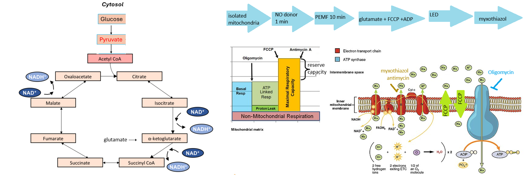
Fig 2 PEMF and state 3 respiration
State 3 respiration is basically that O2 consumption generated by the addition of ADP, that is phosphorylated by complex V as H+ traverse through their electrochemical gradient established by complexes of the electron transport chain. It is complex IV that consumes O2. Electrons and protons, in the form of NADH, come from the tricarboxylic acid cycle. In this case 90 min and 24 hour time points were examined. Pyruvate and malate were added as TCA cycle substrates without an effect. The amino acid glutamate slightly bumped up O2 consumption in response to PEMF. Many amino acids can feed into the TCA cycle as an energy reserve back up. The kicker is that glutamate is indirectly connected to the urea cycle that makes NO. The influence of glutamate is small but statistically significant.
Fig 3 Poisoning liver and muscle homongenates with NO
NO binds to the heme group of complex IV, inhibits electron transport, and O2 consumption. The hypothesis was tested that PEMF can kick NO off complex IV restoring O2 consumption. PEMF did nothing.
Fig 4 Poisoning isolated mitocondria with NO
- State 3, ADP dependent O2 consumption, was increased by PEMF without NO exposure. With NO exposure, there was no difference. This suggests involvement of complex IV in the PEMF effect.
- State 2 respiration is also known as residual O2 consumption. ADP is added but there are no metabolic substrates to generate NADH. There are other enzymes that consume O2 that this post will not go into. Exposure to PEMF greatly increased O2 consumption. NO inhibited the PEMF boost in O2 consumption.
- Respiratory control is the state3/state2 ratio. Obviously one would want most O2 consumption to go to generation of ATP. The larger the better. PEMF boosts this ratio, but only when there is no NO donor.
- When H+ freely traverse the inner membrane via FCCP, O2 consumption is totally uncouple from ATP synthesis and proceeds at a maximum rate. Neither PEMF or NO donors make a difference in this process.
Fig 5, adding myxothiazol a complex III inhibitor
Things start getting more mechanistic in this part of the journey of understanding. According to the authors myxothiazol is a Complex III inhibitor. FCCP was added to abolish the proton gradient.
- Blue light did nothing to change O2 consumption. Poison with NO, blue light slightly increased O2 consumption.
- In the presence of myxothiazol and FCCP, and PEMF, blue light did not increase O2 consumption.
- When an NO donor was added to the mix of FCCP and myxothiazol, blue light increased O2 consumption. The O2 consumption. was still significantly lower than in the absence of NO.
According to Bench Chem, things are a bit more complicated:
“Mechanism of Action
Myxothiazol exerts its antifungal effect by inhibiting the mitochondrial electron transport chain
at Complex III (also known as ubiquinol-cytochrome c reductase).
Binding Site: It binds competitively to the ubiquinol oxidation (Qo) site on cytochrome b, one
of the key subunits of the Complex III.
Inhibition of Electron Transfer: This binding event physically blocks the transfer of electrons
from ubiquinol (Coenzyme Q10, reduced form) to the Rieske iron-sulfur protein (ISP).[2][8]
Disruption of the Q-Cycle: By preventing the oxidation of ubiquinol at the Qo site,
Myxothiazol effectively halts the proton-motive Q-cycle. This cycle is essential for pumping
protons across the inner mitochondrial membrane, which generates the electrochemical
gradient required for ATP synthesis.
Consequences: The immediate consequence is a near-complete blockage of oxygen
consumption and a halt in mitochondrial ATP production, leading to the cessation of cell
growth.”

Wikipedia authors have a few things to say about complex III inhibition that greatly complicates our story.
Fig 6, watching O2 consumption
This is a nice figure that explains a lot. The authors are simply looking at the disappearance of O2.
- Glutamate, malate, and ADP substrates are added. O2 levels very slowly decrease
- Around 8 minutes the NO donor, PEMF, or the control followed by three doses of FCCP to destroy the H+ gradient, faster decrease in O2
- Around 18 minutes the system is exposed to blue light. The rate of O2 consumption is slightly increased.
- When the complex III inhbitor myxothiazol is added, O2 consumption is practically inhibited.
Very interesting study, but…
This study is unique in that it attempts to establish where in the electron transport chain the Hofmag brand of PEMF is acting. It fails to address the usual component of increases in intracellular Ca2+ in homogenates and isolated mitochondria. This is not necessarily a short coming, just an area of further study. Why not measure the effects of the extremely low frequency PEMF devices on complex IV and so on? What about Maghof’s competition. This very interesting study seems o be one of a kind and worthy of follow up. This is the closest study that seems to even come close:
Burlaka A, Selyuk M, Gafurov M, Lukin S, Potaskalova V, Sidorik E. Changes in mitochondrial functioning with electromagnetic radiation of ultra high frequency as revealed by electron paramagnetic resonance methods. Int J Radiat Biol. 2014 May;90(5):357-62.
This is a direct quote of the abstract as the publication is not public access. Ultrahigh frequency was unfortunately not defined in the abstract. Note that there is not indication that the authors were measuring actual O2 consumption in their study.
Abstract
Purpose: To study the effects of electromagnetic radiation (EMR) of ultra high frequency (UHF) in the doses equivalent to the maximal permitted energy load for the staffs of the radar stations on the biochemical processes that occur in the cell organelles.
Materials and methods: Liver, cardiac and aorta tissues from the male rats exposed to non-thermal UHF EMR in pulsed and continuous modes were studied during 28 days after the irradiation by the electron paramagnetic resonance (EPR) methods including a spin trapping of superoxide radicals.
Results: The qualitative and quantitative disturbances in electron transport chain (ETC) of mitochondria are registered. A formation of the iron-nitrosyl complexes of nitric oxide (NO) radicals with the iron-sulphide (FeS) proteins, the decreased activity of FeS-protein N2 of NADH-ubiquinone oxidoreductase complex and flavo-ubisemiquinone growth combined with the increased rates of superoxide production are obtained.
Conclusions: (i) Abnormalities in the mitochondrial ETC of liver and aorta cells are more pronounced for animals radiated in a pulsed mode; (ii) the alterations in the functioning of the mitochondrial ETC cause increase of superoxide radicals generation rate in all samples, formation of cellular hypoxia, and intensification of the oxide-initiated metabolic changes; and (iii) electron paramagnetic resonance methods could be used to track the qualitative and quantitative changes in the mitochondrial ETC caused by the UHF EMR.
Leave a Reply