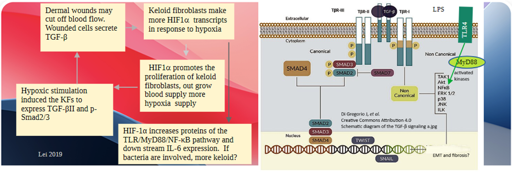
The featured image was adapted from the study of Lei 2019 who addressed the “feed forward” synergy between TGFβ and hypoxia inducible factor alpha (HIFα). The authors were interested in why these scars grow so out of control and exceed the expected margins of the wound. It all has to do with gene transcription triggered by the HIFα transcription factor partner HIFβ. Does red light simply stop this cycle by increasing circulation and oxygenation? What if red light can not only stop fibrosis but also reverse it? We still do not know how, but the answer to this reversal seems to be a collagen active protease called PRss35. Though no clinical trial data was found for keloid scars, an argument is presented that red light might reverse this type of fibrosis.
Keloid and red red light basics
This post proposes that red light might mediate keloid scar removal by a mechanism separate from reactive oxygen species, ATP production, increased blood flow. An introduction is in order to frame this new mechanistic proposal.
HIFα , hypoxia revisited
This image first appeared in the transcranial PEMF post. Our bodies expend an enormous amount of energy in the effort to respond to changes in the environment, in this case, O2. Under normal O2, HIFα gets hydroxyl groups attached to prolines by PHD. Those OH groups serve as an attachment site for the “eat me” ubiquitin tag. A proteosome, which kind of looks like the “Planet Killer” from the first Star Trek series comes along and eats the transcription factor that the cell used a lot of ATP to make. Note that the transcript for growth factor TGFβ is controlled by the HIFα/HIFβ complex.
Note that the growth factor TGFβ is very much part of the response to hypoxia. Surely this explains why keloid growths are out of control and restoring blood flow could dampen that out of control growth. It does not explain the shrinkage of keloids in response to red light.
TGFβ and collagen introduction
How can red light, that aids in wound healing, also lessen the fibrosis that has already occurred? Red light therapy (RLT), we are told, involves production of small amounts of reactive oxygen species. This is a link to aslide show on TGFβ and fibrosis as it relates to cancer. TGFβ signaling can just as easily relate to scars with a lot of fibrosis. TGFβ plays a role in this out of control production of collagen. This post explores a few things about activating transforming growth factor TGFβ that turns on the collagen factory when it escapes the latent state and binds to its receptor.
Note the presence of Furin, a protease that might be part of why red light shrinks keloids. Furin is a protease that cleaves many enzymes into their active form. Wiki authors mentioned matrix metalloproteinase, that is also known as collagenase. Furin may also activate the PRss35 protease shown to be increased 60x in fibroblasts in response to 630 nm red light.
Red Light Home Optimized Total Spectrum Mode for Scars
Red Light Therapy Home offers a product that addresses scars. “For targeted scar treatment, use the Skin & Anti Aging mode on our Total Spectrum devices, which balances intensities to promote collagen without overheating.” All of these device settings are based on peer reviewed research. The company used AI to help formulate claims of close to 3000 peer reviewed studies.
| Channel | Wavelengths | Intensity | wavelength application from Red Light Home |
|---|---|---|---|
| 1: Red | 633nm | 80% | Skin rejuvenation, acne, wound healing |
| 660 nm | Anti-aging, collagen boost, pain relief | ||
| 2: NIR | 810 nm | 20% | Inflammation reduction, deep tissue repair |
| 830 nm | Inflammation reduction, deep tissue repair | ||
| 850 nm | Fat reduction, athletic performance | ||
| 3: Deep NIR | 1064 nm | 0% | Deep tissue pain, skin lesions, brain PBM |
| 4: Blue | 480 nm | ON for first 4 mins | RLTH did not get into anti-bacterial aspects of blue light but this post will mention the TLR4 receptor for bacterial lipopolysaccharide. |
Duration: 10 mins | Pulse: OFF | Beginner Distance: 3+ feet away. Adjust based on scar maturity; recent scars may benefit from more frequent sessions.
Note the collagen boost and wound repair in the 630ish nm red. These two things are sort of opposite keloid shrinkage. RLTH used AI to go through thousands of peer reviewed reports to develop protocols. The PRss35 hypothesis has to take these observations into account.
Prananda 2025, red light for keloid scars
“ Keloids are pathological scars resulting from dysregulated wound healing, marked by excessive deposition of fibrous tissue that extends beyond the original wound margins. In contrast to hypertrophic scars, keloids exhibit continuous growth in the absence of additional trauma”
- Down regulation of VEGF disrupts angiogenesis, thus limiting scar over growth.
- Collagen, types I and III, extend to the extracellular matrix. The Paranda 2025 also discussed different isoforms of collagen, abnormal glycoaminoglycans, matrices so tight that signaling molecules cannot get in, lack of remodeling that involves some collagen degradation, and more.
- Myofibroblasts, arise from TGF-β1–induced differentiation of fibroblasts. As the name suggests, myofibroblasts have contractile properties. Keloid scar formation involves myofibroblasts that arise through TGF-β1–induced differentiation of fibroblasts during wound healing. Other precursor cells may include pericytes and mesenchymal stem cells. In keloids myofibroblasts continue to proliferate and deposit collagen.Red light’s anti-inflammatory effects arise from decreases in IL-6 and TNF-α levels.
- This review covered the usual red light inhibition of TGF-β1 expression somehow via the generation of reactive oxygen species from the mitochondria.
- The myofibroblasts have contractile properties of muscle cells too. It is thought that this contraction allows TGF-β to escape from its LAP cage so that it can elicit the formation of more collagen. See the slide show for a reminder.
- Fig 1 is a cartoon summarizes ways in which RLT promotes wound healing. ATP from activated mitochondria can lead to proliferation via the RAS/Raf/MEK that occurs with uncontrolled proliferation such as cancer. The other arm is reactive oxygen species is the PI3K/Akt pathway that leads to programed cell death, aka apoptosis.
- Macromolecular crowding of extracellular matrix proteins and altered hydration was given brief mention. This concept will become important later.
Note the emphasis on contraction and mechanical things. The macromolecular crowding might feed into osmotic activation of the red light proteases PRss35. Hold the thought of the properties of gelatin prepared with minimal water.
Reactive oxygen species often oxidize amino acid side chains. Cysteines are often targets.
Mamalis 2016a, Red Light Therapy prevents skin fibrosis
- Fig 1 Most fibrosis occurs in the dermis. Sometimes collagen deposition can get out of control RLT returns things to normal.
- Fig 2 shows how the authors surveyed the literature.
- Fig 3 tells us not only about how ROS from the mitochondria affect LAP-TGFβ outside the cell but also what happens when it binds to its receptor. In step 4 of the cartoon, reactive oxygen species have been shown to trigger a conformational change in LAP, thus freeing TGFβ1 from its latency peptide. How then is RLT is good thing (for keloids) if it leads to TGFβ binding to its receptor and producing more collagen? Studies were cited showing RLT leads to ROS that turn on redox sensitive gene transcripts.
This reactive oxygen species (ROS) model has nothing to say about HIFα and O2. Yes, red light returns things to normal as per Fig 1 We are left in the dark as to how red light might shrink keloids. It is just one of the constant skin turn over things? This post says “probably not.”
Mamalis 2016b, 633 nm ± 30 nm red light, fluence on collagen
FlueFluence is the number of particles or the energy per unit area with which a material is irradiated. The material in this case is human dermal fibroblasts on round cell culture dishes. Exposure time was 18 hours.
- Cell counts decrease when going from no exposure to 320 and 640 J/cm2.
- Reactive Oxygen Species (ROS) increase when going from no exposure to 320 and 640 J/cm2.
- Collagen decreases when going from no exposure to 320 and 640 J/cm2.
- Migration speed decreases when going from no exposure to 320 and 640 J/cm2.
These results are extremely excited. What the authors did not do was prove that the ROS were the cause of the decrease in collagen, cell migration, and so on. We do not know for certain that one is causing the other. Some authors use ROS scavengers to prove ROS involvement. Nonetheless, this is still a very good story. ROS will have a home in the new mechanism proposal for red light des.
George 2018, fluence, NIR vs 636 nm red, ROS
Michael Hamblin is a leading expert, if that a grand father, of photo modulation therapy. This particular dermal fibroblast study was conducted when he was a visiting professor at the University of Johannesburg in South Africa. The idea was to expose human dermal fibroblasts to two different wavelengths of light and compare reactive oxygen species, ATP production, cell viability, and so on. The responses to 636 nm vs 825 nm did not track one another as a function of fluence.
The light source
The light source used was a commercially available Omnilux New-U hand held LED array (PhotoTherapeutics, CA). This LED unit has a 4.7 cm × 6.1 cm rectangular aperture and emits visible red light (633 nm ± 30 nm) at a power density of 360.2 W/m2 at room temperature and a distance of 10 mm from the bottom of the tissue culture dish to the LED array.
Summary Fig 7
This figure compares the cell response to 636 nm vs 825nm. Looking at it is like searching for differences in two similar images with slightly different colorings. Here are the similarities.
- We’ve got the usual red and NIR light causing generation of ROS by the mitochondria
- ROS cause the release of Ca2+ from intracellular stores.
- ROS and the ADP/ATP supply modulate adenlyate cyclase which makes the second messenger cyclic AMP, which may modulate two protein kinases, according to this cartoon. Also in the spirit of cAMP we have changes in gene transcription via the cyclic AMP Response Element (CRE) NIR seems to be more effective at increasing ATP production.
- the intensity and the number of fibroblasts stained with ROS dye was higher for fluences above 15 J/cm2 in 825 nm source. ROS may also come from NADPH oxidase. (ACE inhibitors are used to prevent activation of an NADPH family member in our musculature. ) These ROS target growth factor receptor tyrosine kinases that control cell response to growth factors. Which isoform of NADPH oxidase are in fibroblasts? An Internet search lead to Hecker 2009 and Nox4. This particular publication also claims that Nox4 produces H2O2.
- The perplexing thing is that George 2018 did not give the slightest hint on how exciting water molecules would activate NAPH oxidase. Mitochondrial Cox1 has heme groups and so do the NADPH oxidases (Sumimoto 2008) Not only that, but this enzyme family has two heme groups juxtaposed with an FAD and an NADH.
Sometimes the scientific literature can become very confusing when different groups have completely different takes on how things work. What I find interesting is are the subtle differences in 336nm and 825nm. I think the possibility of water being an antenna for NIR an intriguing on and worthy of further exploration.
The oxidation of methionine in LAP, Jobling 2006
“The three mammalian transforming growth factor beta (TGFβ) isoforms are each secreted in a latent complex in which TGFβ homodimers are non-covalently associated with homodimers of their respective pro-peptide called the latency-associated peptide (LAP). Release of TGF-beta from its LAP, called activation, is required for binding of TGFβ to cellular receptors, making extracellular activation a critical regulatory point for TGF-beta bioavailability. Our previous work demonstrated that latent TGFβ1 (LTGF-beta1) is efficiently activated by ionizing radiation in vivo and by reactive oxygen species (ROS) generated by Fenton chemistry in vitro. In the current study, we determined the specific ROS and protein target that render LTGF-TGFβ redox sensitive. First, we compared LTGF-beta1, LTGF-β2 and LTGF-β3 to determine the generality of this mechanism of activation and found that redox-mediated activation is restricted to the LTGF-beta1 isoform. Next, we used scavengers to determine that ROS activation was a function of OH(.) availability, confirming oxidation as the primary mechanism. To identify which partner of the LTGF-β1 complex was functionally modified, each was exposed to ROS and tested for the ability to form a latent complex. Exposure of TGFβ1 did not alter its ability to associate with LAP. Exposing LAP1 to ROS prohibited this phenomenon, while treatment of ROS-exposed LAP1 with a mild reducing agent restored its ability to neutralize TGFβ1 activity. Taken together, these results suggest that ROS-induced oxidation in LAP-TGFβ1 triggers a conformational change that releases TGF-beta1. Using site-specific mutation, we identified a methionine residue at amino acid position 253 unique to LAP-beta1 as critical to ROS-mediated activation. We propose that LTGF-beta1 contains a redox switch centered at methionine 253, which allows LTGF-TGFβ1 to act uniquely as an extracellular sensor of oxidative stress in tissues.”

An image of the N-terminal LAP domain of TGFβ is color coded to show different amino acids. The only cysteines in this domain are the ones involved in disulfide bonds. Unfortunately Met353 is not part of this structure. There are more methionines. This is why Jobling used site directed mutagenesis. We are also led to think that none of the thiols in the active growth factor are involved in his inhibition of the ability to be caged by LAP.
Jobling 2006 used a very strong reactive oxygen species generating system, H2O2 is sufficient to oxidize protein methionines to methionine sulfoxide according to Wikipedia authors; the Jobling 2006 results may also apply to milder ROS from red light therapy. What we need is proof that this methionine can be oxidized in response to red light therapy. Even with this, this does not fully explain how red light can shrink keloids.
water and absorption of red and NIR light
This is a temporary video of what happens when water absorbs red light. This is a cell phone video of several Wikimedia Commons videos of vibrational modes of water. A vibrational mode of water by Evan585619 Creative Commons Attribution-Share Alike 4.0. In watching these water vibrational states, think about the ordered water of George 2018.
The difference between red and NIR is that George and coauthors 2018 claim that water molecules on the surface of the cell absorb in the 825nm NIR range. Wikipedia authors have assembled a good page on water absorption of the electromagnetic spectrum. These authors report absorption of water in the mid infrared region of the spectrum that translates into vibrational states of water. A graph shows an increase in absorbance around 825 nm. Weyer and Lo 2006 also resented a table on water absorbances in the red and NIR. For an explanation of some of the vibrational modes that are IR active, visit this YouTube video. When molecular bonds absorb NIR, they start moving around. According to Sekar 2017 published major IR peaks of DRY collagen. It is an interesting read but rabbit hole diverting us from the main story on how RL and NIR works on keloids. The George 2018 comment on NIR absorption of surface organized water is interesting for those that have made gels with the collagen hydrolysis product know as Gelatin. Trapping of water molecules in the structure was mentioned by Wikipedia authors. An absorption spectra of hydrated collagen in the near infrared part of the electromagnetic spectrum has not been found. The measurements of Sekar 2017 were performed in the dry state between 600-1700 nm on commercially obtained collagen flakes. Their set up was elaborate to control for light scattering, intrinsic fluorescence, and so on.
Water hydrogen bonding to collagen fibrils, one would think, is close to near freezing water. The Prananda 2025 claim that there is macro molecular crowding of collagen in keloid scars drives the hypothesis that bending and stretching of water molecules in this matrix may loosen things up. What happens next? Is there a mechanical force elicited that breaks TGFβ free from its LAP cage? When TGFβ is freed, does it stick around and act in a paracrine fashion, or does it get swept away and go somewhere else? Are there anti-fibrotic signaling molecules lurking around in the collagen mess that are loosened up by red light and NIR?

According to Wikipedia, gelelatin is one of the main ingredients of Gummi Worns. in addition to sugar, flavorings and dyes. Scientific American has a short piece on gelatin, a protease digest of collagen that forms geld upon heating. Nanoscience has some nice scanning electron microscope images of human skin that show gel like structures. These gel like structures are connected to other gel like structures just below the cell membranes via proteins called integrins. Thiago Leal has published a nice slide show on the gel like structures beneath the cell membrane.
The next section presents two publications claiming that red light and osmotic stress induce the expression of a collagen degrading protease. This post could go down all sorts of rabbit holes regarding mechanisms. Let’s keep the concept of Gummy Worms and water trapped in their protein matrix. Doing something to the water so intimately associated to proteins may do things to the proteins themselves.
Red light PRss35 collagen protease transcription
PRss35 was initially dismissed and thought to be an inactive “serine protease” because it had a threonine in its active site. In reality, it requires some special processing to become an active protease. PRss35 includes many of the other aspects of the fibrosis/keloid vicious cycle, water structure, and so on.
Red light induces expression of PRss35, Austin 2021
Austin 2021 used four different human dermal fibroblast cell lines to test the hypothesis that exposure to 633 nm red light for 1, 12, and 24 hours would alter gene expression. Genes were grouped in families and general roles in the cell. Collagen degrading matrix metalloprotease 1 (MMP1) stood out a gene of interest. The differential expression of MMP1 (fold change: 2.36, p-value: 9.86 × 10⁻¹⁴, FDR: 5.02 × 10⁻¹¹ at 24 h) is shown as a representative gene of interest involved in extracellular matrix organization. PRSS35, which produces a serine protease with collagen 1 degrading potential, was found to have greater than 30 fold increased expression at 4, 12, and 24 h after RL treatment, and thus may impart anti-fibrotic effects via collagen degradation
what was done
Human dermal fibroblasts were treated with red light at 633 ± 30 nm. Cells were treated to 320 J/cm2 or 640 J/cm2 (3667 s for 320 J/cm2 and 7334 s for 640 J/cm2) of RL at approximately 34 ˚C. Though cells were not treated with TGFβ1 , down stream signaling molecule transcripts were decreased. A light increase was seen in ROS and a time dependent increase in mmp1 transcripts and translated protein.
results and comments bullet points
- Red light increased ROS.
- To further characterize the potential of red light in fibrosis, differentially expressed genes DEGs in the TGF-β signaling pathway were analyzed.
- Previously work from this group demonstrated that 640 J/cm2 RL decreases SMAD-2 phosphorylation in TGF-β1 induced HDFs within 4 h of exposure. SADs 2/3 translocate to the nucleus to induce the transcription of collagen genes.
- The TGF-β induced, red light SMAD3 at 4, 12, and 24 h post-RL treatment.
- TF analysis confirmed at SMAD3 was likely involved with regulation of cellular activity following red light exposure. Mixed results were seen with red light in regards to pro- and anti-fibrotic SMADs.
- PRSS35 is a supposedly inactive serine protease with a threonine in the active site according to UniProt.
- Wang 2023 found that PRSS35 had trypsin like activity Fig 2 The protease was expressed in E coli, activated by the furin protease and incubated with β-casein. Peptides were sequenced. Supplemental data panel 2f suggests HNF4A binding sites in the PRSS35 promoter.
PRss35 in response to osmotic stress
Sänger 2023 used a 3D culture model and next-generation sequencing to identify genes that are regulated by hyperosmolarity in primary human dermal fibroblasts (HDFs). The most strongly up-regulated gene was pRSS35. The kinase/transcription factor pairs were p38→ATF2 and JNK→NFAT5. Fig 8 summarizes how hyperosmotic stress activates signaling cascades that result in PRss35 transcription and then extracellular matrix degradation. Activated transcription factor 2 (ATF2) is phosphorylated by both JNK and p38 kinases in response to environmental stresses. According to Wiki authors, ATF5 binds to the cAMP response element in the promoters of many genes. Nuclear Factor of Activated T cells 5 (NFAT5) responds to osmotic stress via a protein called Brx that binds to the cell membrane/cytoskeleton junction.

This post deviates from the wound remodeling theme of Sänger 2023. According to the Wiki NFAT5 page, hyperosmotic stress is transmitted to the cytoskeleton where it joins the cell membrane. All sorts of stresses occur here. The osmolyte is sucking water out of the cell membrane. NIR of George 2018 might be doing similar things with “surface associated waters.” See the slide show for a reminder that the collagen extracellular matrix is very much connected to the cytoskeleton.
It doesn’t look like we fully understand how osmotic stress on the cytoskeleton and connected signaling molecules, let alone red light, can activate these kinases that activate transcription factors, that produce transcripts for this collagen degrading protease….Is there some sort of ROS process going on? and Ca2+ signaling?
Clinical trials
BCli
NCT02630303 The goal of this study is to establish the safety of high fluence LED-RL from 160 J/cm2 up to 640 J/cm2 in healthy subjects. The hypothesis is that high fluence LED-RL phototherapy is safe in human skin. The results were not posted on clinicaltrials.gov or published in the peer reviewed literature. They were no doubt used to justify the safety of further clinical trials.
NCT03795116 Light Emitting Diode-Red Light (LED-RL) Phototherapy for Skin Scarring Prevention
Completed WITH RESULTS The results of this 633 nm red light and the sham on face lift surgery scars were published in a peer reviewed study ikurtti (2021)
“There were no significant differences in scar pliability between treated and control scars. At certain fluences, treated scars showed greater improvements in observer rating and scar pliability, reflected by greater reductions in induration, from baseline to 6 months compared to control scars. Treatment-site adverse events included blistering (n=2) and swelling (n=1), which were mild and resolved without sequelae.” See the graphical summary.
So what if red and NIR light can reverse the fibrosis in keloid scars? It could reverse fibrosis in other disease conditions that are more than cosmetic? A clinical trial establishing efficacy could have broader implications. A mechanism of inducing this fantom serine (threonine) protease PRss35 would be nice.
References
- Austin E, Koo E, Merleev A, Torre D, Marusina A, Luxardi G, Mamalis A, Isseroff RR, Ma’ayan A, Maverakis E, Jagdeo J. (2021) Transcriptome analysis of human dermal fibroblasts following red light phototherapy. Sci Rep. 2021 Apr 1;11(1):7315. PMC free paper
- George S, Hamblin MR, Abrahamse H. (2018) Effect of red light and near infrared laser on the generation of reactive oxygen species in primary dermal fibroblasts. J Photochem Photobiol B. 2018 Nov;188:60-68. PMC free paper
- Hecker L, Vittal R, Jones T, Jagirdar R, Luckhardt TR, Horowitz JC, Pennathur S, Martinez FJ, Thannickal VJ. (2009) NADPH oxidase-4 mediates myofibroblast activation and fibrogenic responses to lung injury. Nat Med. 2009 Sep;15(9):1077-81. PMC free paper
- Jobling MF, Mott JD, Finnegan MT, Jurukovski V, Erickson AC, Walian PJ, Taylor SE, Ledbetter S, Lawrence CM, Rifkin DB, Barcellos-Hoff MH. (2006) Isoform-specific activation of latent transforming growth factor beta (LTGF-beta) by reactive oxygen species. Radiat Res. 2006 Dec;166(6):839-48. PubMed
- Kurtti A, Nguyen JK, Weedon J, Mamalis A, Lai Y, Masub N, Geisler A, Siegel DM, Jagdeo JR. (2021) Light emitting diode-red light for reduction of post-surgical scarring: Results from a dose-ranging, split-face, randomized controlled trial. J Biophotonics. 2021 Jul;14(7):e202100073. PMC free paper
- Lei R, Li J, Liu F, Li W, Zhang S, Wang Y, Chu X, Xu J. (2019) HIF-1α promotes the keloid development through the activation of TGF-β/Smad and TLR4/MyD88/NF-κB pathways. Cell Cycle. 2019 Dec;18(23):3239-3250. PMC free paper
- Mamalis A, Siegel D, Jagdeo J. (2016a) Visible Red Light Emitting Diode Photobiomodulation for Skin Fibrosis: Key Molecular Pathways. Curr Dermatol Rep. 2016;5:121-128. PMC free paper
- Mamalis A, Koo E, Garcha M, Murphy WJ, Isseroff RR, Jagdeo J. (2016b) High fluence light emitting diode-generated red light modulates characteristics associated with skin fibrosis. J Biophotonics. 2016 Dec;9(11-12):1167-1179. PMC free paper
- Prananda AT, Syahputra RA. (2025) Photobiomodulation therapy in keloid management: a comprehensive review. Front Med (Lausanne). 2025 Jul 9;12:1550662. PMC free paper
- Sänger CS, Cernakova M, Wietecha MS, Garau Paganella L, Labouesse C, Dudaryeva OY, Roubaty C, Stumpe M, Mazza E, Tibbitt MW, Dengjel J, Werner S. (2023) Serine protease 35 regulates the fibroblast matrisome in response to hyperosmotic stress. Sci Adv. 2023 Sep;9(35):eadh9219. PMC free paper
- Sekar SK, Bargigia I, Mora AD, Taroni P, Ruggeri A, Tosi A, Pifferi A, Farina A. (2017) Diffuse optical characterization of collagen absorption from 500 to 1700 nm. J Biomed Opt. 2017 Jan 1;22(1):15006. free paper
- Sumimoto H. (2008) Structure, regulation and evolution of Nox-family NADPH oxidases that produce reactive oxygen species. FEBS J. 2008 Jul;275(13):3249-77. free paper
- Wang T, Zhou Y, Zhou Z, Zhang P, Yan R, Sun L, Ma W, Zhang T, Shen S, Liu H, Lu H, Ye L, Feng J, Chen Z, Zhong X, Wu G, Cai Y, Jia W, Gao P, Zhang H. (2023) Secreted protease PRSS35 suppresses hepatocellular carcinoma by disabling CXCL2-mediated neutrophil extracellular traps. Nat Commun. 2023 Mar 18;14(1):1513. PMC free paper
- Weyer LG and Lo SC (2006) Spectra–Structure Correlations in the Near-infrared. Handbook of Vibrational Spectroscopy, Online © 2006 John Wiley & Sons, Ltd. This article was published in the Handbook of Vibrational Spectroscopy in 2006 by John Wiley & Sons, Ltd free paper
Leave a Reply