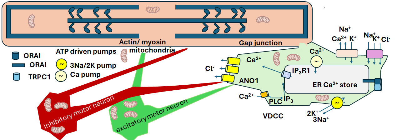
Interstitial Cells of Cajal (ICC) are pace keepers of our colonic contractions. That have the voltage gated Ca2+ channels and Ca2+ chloride channels. They stoe Ca2+ too! We cannot forget that Ca2+ causes smooth muscle of our colon to contract. When damaged, constipation ensues. Moving magnetic fields can also move charged particles from our microbiome activities, good and bad. This post is devoted to the notion that PEMF can get things moving through our guts in multiple ways. Just be careful with crazy, insane, long exposures!
ICC, pediatric gastrointestinal motility, Friedmacher &Rolle 2023
“Over the years, various gastrointestinal motility disorders have been described in infants and young children in which symptoms of functional bowel obstruction arise from ICC-related neuromuscular dysfunction of the colon and rectum. ”
According to this review Interstitial cells of Cajal (ICC)
- ICC are pacemaker cells that propagate electrical slow waves to gastrointestinal smooth muscle cells. This is a direct coupling.
- ICC mediate both inhibitory and excitatory motor neurotransmission from the enteric nervous system. This goes with the featured image.
- Mechanical stretch sensing ion channels are involved in ICC activity. Could this be via Stim1, Orai, and Trpv1?
- ICC form networks with intramuscular terminals of vagal afferents and may also have a role in afferent signaling. Afferents being neurons that take messages back to the brain.
While this review has little “electrical” information of the ICC, it presents an absolutely stunning array of fluorescent immuno cytochemistry images. These do a fantastic job of moving us past cartoons that do not show us how crowded biological molecules are in situ.
ICC, kids with refractory constipation, Yuan 2023
Yuan 2023 is only available for free in abstract form. They compared children with and without refractory constipation with colonic manometry and just a few full thickness colonic biopsies. The intestinal biomes were also studied. Abnormal ICC morphology was observed, microbiota disruptions, and changes in waves of muscle contraction. After looking at the basic physiology of how ICC work, this post will present some links to copyright images and explore how the microbiome contributes to the circuitry. Let’s first look at what ICC do based on what proteins they express.
The protein tool box of ICC tell us what they do, Lee 2017
Many excellent reviews and studies have been published on the work of Interstitial cells of Cajal (ICC). Lee 2017 conducted a massive ICC sequencing study of isolates from the jejunum (JICC) and the colon (CICC). I’ve pulled together two tables of ion channels. The first table presents the most abundant ion channel transcripts. The second table pulls channels mentioned in the text of Lee 2017, other posts on this website, and other pblications mentioned in this post. TFPKM stands for FPKM, or Fragments Per Kilobase of transcript per Million mapped reads. There are 567 “ion channels” in the list.
| gene | Expression (FPKM) | |||
| Symbol | Full name | Activity | JICC | CICC |
| Ano1 | anoctamin 1, Ca2+ activated Cl– channel | most sources say depolarization | 702.72 | 1,068.03 |
| Atp2a2 | ATPase, Ca2+ transporting, cardiac muscle, slow twitch 2 | returns Ca2+ to intracellular stores | 133.31 | 847.63 |
| Atp2a3 | ATPase, Ca2+ transporting, ubiquitous | returns Ca2+ to intracellular stores | 674.42 | 657.88 |
| Slc12a2 | Na/K/Cl cotransporter | water follows ions, volume regulation | 334.13 | 527.06 |
| Slc24a3 | (Na+/K+/Ca2+ exchanger), | Ca2+(out) + K+(out) + 4 Na+(in) = Ca2+(in) + K+(in) + 4 Na+(out) | 103.63 | 353.56 |
| Atp5b | ATP synthase, H+ transporting mitochondrial F1 complex, beta subunit | makes ATP | 766.27 | 322.59 |
| Atp5a1 | ATP synthase, H+ transporting, mitochondrial F1 complex, alpha subunit 1 | makes ATP | 403.89 | 221.20 |
| Atp6v0c | ATPase, H+ transporting, lysosomal V0 subunit C | helps acidify lysosome | 197.96 | 220.34 |
| Atp5g3 | ATP synthase, H+ transporting, mitochondrial F0 complex, subunit C3 (subunit 9) | makes ATP | 463.93 | 190.81 |
| Atp1a1 | ATPase, Na+/K+ transporting, alpha 1 polypeptide | establishes membrane potential | 502.26 | 169.97 |
| Atp5j | ATP synthase, H+ transporting, mitochondrial F0 complex, subunit F | makes ATP | 248.47 | 150.67 |
| Atp5l | ATP synthase, H+ transporting, mitochondrial F0 complex, subunit g | makes ATP | 275.02 | 122.53 |
| Atp5k | ATP synthase, H+ transporting, mitochondrial F1F0 complex, subunit e | makes ATP | 324.79 | 121.62 |
| Atp5h | ATP synthase, H+ transporting, mitochondrial F0 complex, subunit d | makes ATP | 247.55 | 115.59 |
| Atp1a2 | ATPase, Na+/K+ transporting, alpha 2 polypeptide | establishes membrane potential | 194.41 | 113.84 |
| Vdac1 | voltage-dependent anion channel 1 | mitochondria outer membrane, cell death | 210.23 | 113.33 |
The expression data spreadsheet of Lee 2024 was searched for genes mentioned in the text. Only ion channels with a score of 10 or higher are shown. While the ryanodine receptor is expressed in very small amounts in ICC, it is not included in the featured image cartoon.
| Gene | Function | Expression (FPKM) | |||
| Symbol | Full name | Activity | Ion | JICC | CICC |
| Cacna2d1 | calcium channel, voltage-dependent, alpha2/delta subunit 1 | VDCC | Ca2+ | 25.51 | 30.41 |
| Cacna1c | calcium channel, voltage-dependent, L type, alpha 1C subunit | VDCC | Ca2+ | 13.13 | 10.97 |
| Cacna1a | calcium channel, voltage-dependent, P/Q type, alpha 1A subunit | VDCC | Ca2+ | 16.57 | 3.66 |
| Itpr1 | inositol 1,4,5-trisphosphate receptor 1, a channel | Ca2+release from ER | Ca2+ | 25.39 | 52.19 |
| Stim1 | stromal interaction molecule 1 | connects Ca2+ deleted ER to ORA Ca2+ | Ca2+ | 31.56 | 39.79 |
| Pkd1 | polycystic kidney disease 1 homolog | Channel, details lacking | Ca2+ | 72.35 | 98.22 |
| Tmem66 | transmembrane protein 66 | inhibits STIM/ORAI 1 | Ca2+ | 65.89 | 66.72 |
| Gas6 | growth arrest specific 6 | binds Ca2+, few details | Ca2+ | 60.04 | 42.69 |
| Trpc1 | transient receptor potential cation channel, subfamily C, member 1 | PEMF channel | Ca2+ | 24.46 | 17.00 |
| Orai1 | ORAI calcium release-activated calcium modulator 1 | Channel, connected by STIM1 to ER | Ca2+ | 14.07 | 29.55 |
ICES M1 PEMF and ICC rhythms?
Unlike many commercial PEMF devices available to biohackers, the Micropulse M1 device allows the user some ability to set the frequency. We’ve got two rhythms in these pace maker cells. One is the depolarization and smooth muscle contraction. The other is Ca2+ sparks.
ICC slow waves Hwang 2009
In a previous study Hwang 2009 studied the effect of Cl– channel blockers aimed at ANO1 were used to test the hypothesis that ANO1 splice variants are responsible for pacemaker activity in the mouse, monkey, and human gastrointestinal tract. Nifumic acid was used to block Cl– channels.
| figure | species | location | frequency cycle / min | duration, sec | IC50 μM |
|---|---|---|---|---|---|
| Fig 3 | mouse | antrum | 3.2 | 5 | 5.43± 0.3 |
| Fig 4 | monkey | antrum | 3 | ~2.7 | 3.59 ±1.4 |
| Fig 5 | mouse | small intestine | 32 | ~0.8 | 150±3.9 |
| Fig 6 | monkey | small intestine | 12 | 2.5 | 160 ± 2.2 |
| Fig 7 | human | small intestine | ~11 | 5? |
The motivated reader is invited to view other figures in the public access publication. This cartoon from López-Pingarrón 2023 illustrates the electrical coupling between ICC, neurons, and smooth muscle. It is one of many mages that inspired the featured image of this post.
Baker 2021 Ca2+ transients
This group used complicated fluorescent imaging techniques to view time dependent changes extracellular Ca2+ changes. The popular culture of PEMF is that it opens voltage gated Ca2+ channels. In this case a quote from the abstract will be enough.
Interstitial cells of Cajal (ICC) generate pacemaker activity responsible for phasic contractions in colonic segmentation and peristalsis. ICC along the submucosal border (ICC-SM) contribute to mixing and more complex patterns of colonic motility. We show the complex patterns of Ca2+ signaling in ICC-SM and the relationship between ICC-SM Ca2+ transients and activation of smooth muscle cells (SMCs) using optogenetic tools. ICC-SM displayed rhythmic firing of Ca2+transients ~ 15 cpm and paced adjacent SMCs. The majority of spontaneous activity occurred in regular Ca2+ transients clusters (CTCs) that propagated through the network. CTCs were organized and dependent upon Ca2+ entry through voltage-dependent Ca2+ conductances, L- and T-type Ca2+ channels. Removal of Ca2+ from the external solution CTCs. Ca2+ release mechanisms reduced the duration and amplitude of Ca2+ transients but did not block CTCs. These data reveal how colonic pacemaker ICC-SM exhibit complex Ca2+-firing patterns and drive smooth muscle activity and overall colonic contractions
video 1 is an amazing video showing Ca2+ increases in ICC and SM at the same time. The viewer may even see changes in the time course of Micro-Pulse devices. Video 2 shows high spacial resolution of Ca2+. It is best to go directly to the paper for this one.
- Fig 1 Focus on Panels A&B red fluorescent staining for ICC marker kit. The fluorescence disappears (panel C) when the submucosa is stripped away. This is the part that is exposed to the microbiome.
- Fig 2 is one of those beauty of nature fluorescent images that show the close spacial proximity of smooth muscle cells and ICC. It also shows temporal displacement of Ca2+ transients. Does PEMF change conductance of Ca2+ through ICC-SM gap junctions?
- Fig 3 some spacial temporal stuff
- Fig 4 Near by sub mucosal IC dihydroC talk to each other.
- Fig 5 shows high time resolution at multiple firing sites leaving open the possibility that some of the Micropulse M1 settings might be in sync.
- Fig 6… average percentages of firing sites during a Ca2+ wave. Values are calculated for 1 s and plotted in 85 ms bins
- Fig 7 Ca2+ outside the cell matters. What does this mean if PEMF can get extracellular Ca2+ moving?
- Fig 8, ICC and SM cells were sorted successfully sorting Kit/Ano1 ICC from myosin heavy chain smooth muscle. L-Type Ca2+ channels (Cacna1c and Cacna1d) T-type Ca2+ channels ( Cacna1h) and Store‐operated Ca2+ entry (SOCE) channels (Orai1 and Orai2) were found in ICC Cacna1d and Orai1&2 were more in ICC and smooth muscle cells.
- Fig 9 L-type Ca2+ channel blocker
- Fig 10 CaV3, (T-type) Ca2+ channel blockers
- Fig 11 pinacidil, ATP gated K+ channel blocker; nicardipine dihydropyridine, Ca2+ channel blocker; NNC 55–0396, T-type Ca2+ blocker in combination.
- Fig 12, Thapsigargin is an inhibitor of SERCA, the Ca2+ endoplasmic/sarcoplasmicCa2+ ATPase returnes Ca2+ to intracellular stores is part of the process.
- Fig 13 The ryanodine receptor, TRPV1-3, and and the IP3 receptor Ca2+ channel are a small part of the Ca2+ mobilization picture.
- Fig 14 Store Operated Ca2+ is involved
- Fig 15, ⭐⭐⭐Tying all of this together in a two cartoons.

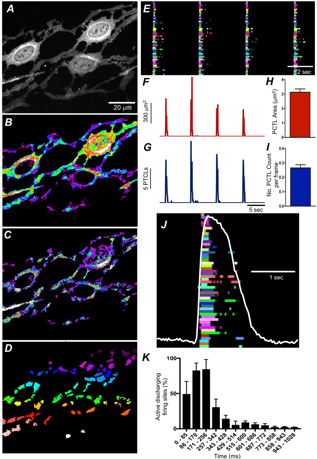
What this means for contracting the colon?

Lee 2017 suggests that ICC can be a buffer for Ca2+ as well as electrical control of the smooth muscle cell membrane potential by way of Ano1. Membrane potential is another way of letting Ca2+ into the smooth muscle cell.

Baker 2021 pulled in acto-myosin contraction of smooth muscle in a Fig 15 supp cartoon. It is time to abandon all hope that PEMF effects only one of the many ion channels that regulate smooth muscle tone. There are many transporters and channels of Lee 2017 that were not explored.
Kaszuba-Zwoińska is another great review on this topic.
The little brain and the microbiome Sharkey and Mawe 2023
The journey up to this point (Baker, Hwang, Lee) has been there are a massive number of ion channels and transporters in the sub mucosal ICC. Sub mucosal means they are in close proximity to the mcirobiome and all the small molecules produced by the microbiome.
Fig 1 Schematic overview of the gut-brain-microbiota axis in micro circuits.
This is a really nice image that also includes spinal cord motor neuron innervation of the gut. The mcrobiome communicates with epithelial cells, including enteroendocrine, chemical sensing tuft cells, and immune cells in the lamina propria that relay messages with enteric nerves, enteric glia to modulate the body’s response to the lumen environment. Mention is given to CNS reflexes. The primary virtue of this cartoon of this cartoon is that it gives an overview of the cell types in a less crowded way.
Fig 2 The myenteric plexus is a very crowded place
Sharkey and Mawe have a fluorescent immunohistochemistry image of neurons (labeled with ant-HuC/D, blue) and glia (labeled with anti-S100B, pink). This figure also shows a cartoon of the sub mucosal plexus of which we’ve just been covering the ICC and smooth muscle cell Ca2+ sharing. The picture is becoming much more complicated just in the submocal layer.
For the PEMF hack a take home message is that things are really crowded in these parts of our bodies. There is always the possibility that PEMF works by stirring up stagnant charged particles in these regions.
Fig. 3 Chemical and mechanical stimulation
Sharkey and Mawe give us the connection between the microbiome and the ICC even though they do not appear in this cartoon. Chemically and mechanically activated enteroendocrine cells form synaptic connections with intrinsic primary afferent neurons. There are also immune cells floating around in the sub mucosa.
For the PEMF hack, just stirring up stagnant chemicals or gentle movement of charged particles might be part of the process.
Fig4 moving poops requires relaxation and contraction of smooth muscle
The text of this figure discusses neurotransmitters and such. “Upon a chemical or mechanical luminal stimulus, intrinsic primary afferent neurons cause the activation of ascending interneurons that synapse with excitatory motor neurons evoking an oral contraction, whereas the activation of descending interneurons leads to the excitation of inhibitory motor neurons eliciting a relaxation anally. ” Neurotransmitters include: nitric oxide, substance P, acetyl choline, VIP, and purines.
Many PEMF hacks like to think that PEMF opens up voltage gated Ca2+ channels, which We have covered in the previous section. Added neurons greatly complicates matters. Surely Faraday’s Law of Induction stirring of these neurotransmitters may change things. If a poop gets stuck, getting it unstuck is a complicated matter.
Fig 5 bile acids, LPS, short and long chain fatty acids
and associated text reiterates the importance of enteroendocrine cells and intrinsic primary afferents. Toll like receptors (TLR) and the the noxious chemical receptor Ca2+ channel TRPA1 are added to the image. Also added are free fatty acid receptors (FFAR). TLR4,in particular, responds to lipopolysaccharide of Gram negative bacteria. Pathogen Associated Molecular Pattern (PAMP) is common to TLR. The aryl hydrocarbon receptor (AHR) transcription factor appeared in the text but not the cartoon. TGR5 is the bile acid receptor
This particular cartoon has a nice layer of mucus in it. Wikipedia authors mention the polyelectrolyte properties of mucus that must include counter ions. Just something to thing about when it comes to Faraday’s law of Induction.
Fig 6 Immune cells are in communication with the Little Brain
This figure does a nice job of summarizing immune cells, neurotransmitters, and cytokines of the sub mucosa. Intrinsic and extrinsic primary afferents commutate with deeper layers of neurons. Afferents carry sensory information from the periphery to the CNS. Intrinsic and extrinsic refer to where the cell bodies of the neurons are located.
This figure presents an assortment of interleukins, cytokines released by cells of the immune system.
It would seem that interleukins seem to influence the health of ICC.
Fig 7 Goblet cells release mucus
This is something to think about when applying any kind of stimulus to teh GI track.
Fig 8 CNS circuits
CNS input is from the spinal cord as well as the vagus nerve. There is no direct CNS connection with muscle, these authors claim, but rather the enteric nervous system. This is a phenomenal figure for PEMF hacks who have tried, sometimes successfully, to get things moving by stimulating the vagus.
Fig 9 Enteroendocrine cells
Enterochromaffin cells and tuft cells are regulated by the microbiota. Tuft cells respond to microbial signals via succinate (SuncR1) and taste (Tas1R/Tas2R) receptors and release acetylcholine (ACh) and immune mediators [IL-25 and leukotriene C4 (LTC4)] to regulate neuroimmune signaling in the lamina propria…. also response to bile acids, short chain fatty acids….Piezo 1 and 2 are stretch sensitive Ca2+ channels. Perhaps they signal the presence of a stool tha is not that compressible.
Fig 10..Microbiomes can be bad influences, consult the text!
With terms like “reactive gliosis” and “neuronal cell death” this figure makes it clear that it is impossible to understand everything. Even if one brand of PEMF or another elicits contraction via voltage gated Ca2+ channels, movement of Ca2+ between ICC and smooth muscle cells, or whatever, it may not cure underlying issues with the microbiome and toxic effects.
Redox reactions and ICC, and killing with PEMF
This section blends what the López-Pingarrón 2023 review says about oxidative stress and ICC and the Kaszuba-Zwoińska 2005 PEMF studies showing ICC killing after prolonged exposure.
quote from López-Pingarrón 2023
Generation of reactive oxygen and nitrogen species (ROS and RNS). ROS are produced during the normal aerobic cell metabolism. Superoxide anion (O2•−) is formed from oxygen (O2) mainly in the mitochondria. O2•− either reacts rapidly with nitric oxide (•NO) to produce toxic peroxynitrite (ONOO−) or is catalyzed by superoxide dismutase (SOD) to generate hydrogen peroxide (H2O2), which can be neutralized by catalase or glutathione peroxidase (GPx). In presence of transition metal ions, such as ferrous ion, H2O2 forms hydroxyl radicals (•OH) by the Fenton reaction. GR: glutathione reductase; GSH: reduced glutathione; GSSG: oxidized glutathione; NADP: nicotinamide adenine dinucleotide phosphate; NADPH: reduced NADP; NOS: nitric oxide synthase.” Link to figure
Long term PEMF exposure
- Kaszuba-Zwoińska exposed rats, in groups of four, to PEMF for four weeks. An equal number of non-exposed rats were used as controls.
- Unlike QuantumTx’s downfield PEMF, the magnetic field generated by coil was directed vertically with a sinusoidal 10 kHz frequency.
- The cages sat in rectangular coils.
- The authors claimed that magnetic fields of such frequencies are commonly generated by audio equipment.
- This frequency is too high to depolarize autonomic fibers, heat the tissue, or other non-disclosed effects.
- Wave bursts with a duration time of 30ms strength of 300µT and frequency of 1Hz
- were generated. The frequency of the RF bursts is on line with the fastest slow waves.
- After each period of exposition tissue samples from both groups were excised from the stomach, duodenum and colon. The tissue sectionsfrom both exposed and unexposed rats were immunohistochemically stained with polyclonal anti-c-kit that the Kaszuba-Zwoińska used as a marker for ICC.
| PEMF exposure | 1 week | 2 weeks | 3 weeks | 4 weeks |
| Duodenum | 63.0± 20.9 * | 32.8 ±9.4 * | 21.1 ± (7.6) * | 23.4 ± 9.5 * |
| Proximal colon | 88.3 ±27.1 | 44.1± 12.1 * | 33.7 ± 6.4 * | 33.7 ± 12.8 * |
| Fundus | 40.1 ±22.7 * | 27.0 ±18.0 * | 18.1 ± 6.6 * | 12.2 ± 9.8 * |
| Pylorus | 100.0 ±10.8 | 93.3 ±10.0 | 99.1 ± 26.9 | 83.3 ± 32.4 |
| Pacemaker | 58.1 ± 27.8 | 78.7 ±16.9 | 57.5 ± 26.8 | 36.7 ± 20. |
able 1. Percentage [%] of ICCs present in the rat’s gastrointestinal tract upon different periods of exposure to PEMF in comparison to control group. Results expressed as mean ± SD.
In a second publication the same year, Kaszuba-Zwoińska 2005, PEMF was shown to generate apoptosis in ICC in a dose dependent as revealed by immunostaining with antibody against proapoptic protein Bax. Bax increases the opening of, the mitochondrial voltage-dependent anion channel (VDAC), which leads to the loss in membrane potential and the release of mitochondrial cytochrome C. Recall that Lee 2017 demonstrated large representation of CDAC mRNA in rat jejunal and colonic ICC.
Lorentzian Force mixing, that’s all?
The Interstitial Cells of Cajal are important players in constipation (Friedmacher 2023, López-Pingarró 2023, Yuan 2023). Transcriptomics reveals that Ca2+ is an important component of ICC functioning. The ICC Ca2+ gated Cl– channel Ano1 is an important part of the slow wave of our colons. (Hwang 2009). The movement of Ca2+ in ICC involves multiple channels (Salah 2021) The Sharkey and Mawe review forces us to thing about the rest of enteric nervous system and the microbiome. Many PEMF hacks are convinced that PEMF works by pushing Ca2+ through voltage gated Ca2+ channels. There are a bunch of them. (Lee, Baker, etc_) Perhaps PEMF just stirs charged paricles up! Wikipedia authors have written extensively on the physics of moving charges creating magnetic fields, the paths taken of moving charges when then encounter magnetic fields, and so on. Perhaps PEMF is a moving electromagnetic field that disperses charged particles doing harm or just in need of being someplace else to do good. Prolonged exposure can do harm (Kaszuba-Zwoińska 2004)

ICC slow waves are keeping pace all over our GI tracts. The Micro-Pulse 1 Hz frequency is still faster than the slow waves even in the stomach. It is a place to start self experimenting. All the mechano-sensors and chemical sensors will be telling those pace maker ICC different stuff. Let your body tell you and don’t over expose!
References
- Baker SA, Leigh WA, Del Valle G, De Yturriaga IF, Ward SM, Cobine CA, Drumm BT, Sanders KM. (2021) Ca2+ signaling driving pacemaker activity in submucosal interstitial cells of Cajal in the murine colon eLife 10:e64099 free paper
- Friedmacher F, Rolle U. (2023) Interstitial cells of Cajal: clinical relevance in pediatric gastrointestinal motility disorders. Pediatr Surg Int. 2023 Apr 27;39(1):188. PMC free paper
- Huizinga JD, Hussain A, Chen JH. (2021) Interstitial cells of Cajal and human colon motility in health and disease. Am J Physiol Gastrointest Liver Physiol. 2021 Nov 1;321(5):G552-G575. free paper
- Hwang SJ, Blair PJ, Britton FC, O’Driscoll KE, Hennig G, Bayguinov YR, Rock JR, Harfe BD, Sanders KM, Ward SM. (2009) Expression of anoctamin 1/TMEM16A by interstitial cells of Cajal is fundamental for slow wave activity in gastrointestinal muscles. J Physiol. 2009 Oct 15;587(Pt 20):4887-904. PMC free paper
- Kaszuba-Zwoińska J, Ziomber A, Gil K, Bugajski A, Zaraska W, Thor P. (2005) Pulsating electromagnetic field induces apoptosis of rat’s bowel Cajal’s cells. Folia Med Cracov. 2005;46(3-4):87-95. PMID: 17252991.
- Kaszuba-Zwoińska J, Gil K, Ziomber A, Zaraska W, Pawlicki R, Królczyk G, Matyja A, Thor PJ. (2005) Loss of interstitial cells of Cajal after pulsating electromagnetic field (PEMF) in gastrointestinal tract of the rats. J Physiol Pharmacol. 2005 Sep;56(3):421-32. free paper
- Lee MY, Ha SE, Park C, Park PJ, Fuchs R, Wei L, Jorgensen BG, Redelman D, Ward SM, Sanders KM, Ro S. (2017)Transcriptome of interstitial cells of Cajal reveals unique and selective gene signatures. PLoS One. 2017 Apr 20;12(4):e0176031. PMC free paper.
- López-Pingarrón L, Almeida H, Soria-Aznar M, Reyes-Gonzales MC, Rodríguez-Moratinos AB, Muñoz-Hoyos A, García JJ.(2023) Interstitial Cells of Cajal and Enteric Nervous System in Gastrointestinal and Neurological Pathology, Relation to Oxidative Stress. Curr Issues Mol Biol. 2023 Apr 18;45(4):3552-3572. PMC free paper
- Sharkey KA, Mawe GM. (2023) The enteric nervous system. Physiol Rev. 2023 Apr 1;103(2):1487-1564. PMC free paper
- Yuan Y, Lu Y, Zhang Z, Cheng W, Yan K, Zheng Y, Jin Y, Liu Z. (2023) Characteristics of the Cajal interstitial cells and intestinal microbiota in children with refractory constipation. Microb Pathog. 2023 Nov;184:106373. 10.1016/j.micpath.2023.106373. PubMed
Leave a Reply