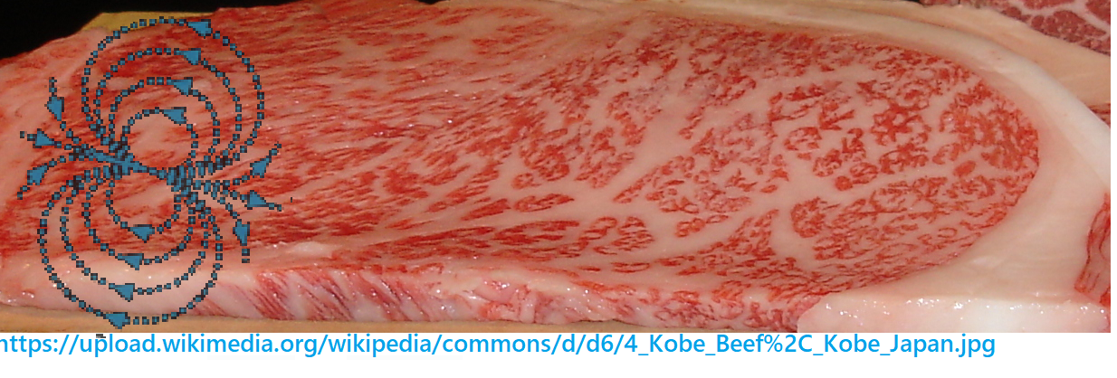
. QuantumTx is a company that uses PEMF to enhance muscle health in the elderly. QuanumTx PEMF affects skeletal muscle and white adipose tissue. The featured image illustrates that skeletal muscle has fat and myocytes. The featured image also suggests an orientation specificity of the magnetic field called “up” and “down”, down being the most effective. And the most important thing, myocytes secrete vesicle that are likely to interact with adipocytes.
The QuantumTx PEMF waveform
The ELF-PEMF (extremely low frequency – pulsed magnetic field) delivery system has been described previously.

For the purposes of this study a barrage of magnetic pulses of 6 ms duration was applied at a repetition rate of 15 Hz and at flux densities between 1–4 mT. Each 6 ms burst consisted of a series of 20 consecutive asymmetric pulses of 150 µs on and 30 µs off duration with an approximate rise time of 17 T/s. All of this comes out to about 5 kHz pulses in between radio and very low regions of the electromagnetic spectrum. See previous post for calculations.

15 Hz, 1.5 mT, less exposure is more [1]
This study fine tuned the timing and field strength of 15 Hz PEMF on mesenchymal stem cells (MSC) chondrogenic differentiation, i.e. cartilage formation. Three markers were examined:
- collagen 2
- Sox9,
- agggregan.
Different duration and field intensity combinations of 15Hz PEMF increased or decreased the mRNA transcripts of these proteins associated with chondrocyte differentiation. This study found that less (10 minutes) is more effective at low intensity (2 mT) exposure to the standard waveform. More exposure diminished chondrogenic outcome. This was attributed to calcium entry after the initial induction. Transient receptor potential (TRP) channels appear to be mechanistic targets. Blocking calcium entry during the repeated PEMF exposure with EGTA or TRP channel antagonists influenced the response to PEMF. The most curious thing is that the optimized combination of duration and magnetic field intensity for differentiation transcripts also yielded the maximum transcripts of TRPA1 and TRPV4.
As an aside, Singh 2002 demonstrated TRPC1channel activity feedback inhibition by Ca2+-calmodulin binding to a domain in the C terminus of TrpC1.
A PEMF mito hormesis mouse study [2]
As one dose of 0.5 to 2 mT Signature PEMF for 10 minutes was optimal for cells, mice were subjected to this dose once on Sundays. Another group of female mice were subjected to intense exercise on Mondays, Wednesdays, and Fridays.
summary of figures and table [2]
- PEMF increased transcripts of PGC–1α, TRPC1, CDC36, myoG, and IGF1. Insulin was decreased whereas free fatty acids, triglycerides, and cholesterol were unchanged.
- PEMF and exercise increased slow twitch fiber promoting transcription factor Pgc-1α transcripts in the slow twitch soleus but decreased them in the extensor digitorum longus (EDL) This was figure 2. Table 2 compared mitohormonic transcripts with ANOVA and t-tests. Only Pgc-1α was significantly increased by pooled PEMF vs pooled control (p=0.03). T-test follow up demonstrated differences between PEMF+ exercise and control/sedentary (p=0.03) and PEMF and no PEMF/exercise (p=0.02). These results suggest that the main targets of PEMF lie elsewhere.
- Brown adipose tissue is brown due to fat burning mitochondria. Figure 3 and Table 3 compared brown and white fat transcripts. 2way ANOVA revealed a PEMF induced increase in Pgc-1α transcripts in brown and white adipose tissue. Many other significant effects of PEMF and exercise were observed in adipose tissues. The only PEMF/exercise interaction in white adipose tissue was seen with the cytochrome C oxidase subunit Cox7a1 transcript. The only significant PEMF/exercise interaction in brown adipose was in reduction of serum leptin.
- Figure 4 is a “role” call of mitohormetic gene transcripts that play a role in mitochondria energetics and metabolism. Some transcripts decreased with PEMF alone while others increased with PEMF alone. PEMF did not have as much positive influence on exercising mice as did PEMF did on sedentary mice. PPARα was increased by about 5x by PEMF in sedentary mice. Nrf1 was increased by PEMF in sedentary mice around 2-3x. There was a lot of variability tin the response! In general, PEMF leveled the mitohormetic transcripts in the sedentary mice to levels found in the exercising mice ± PEMF. There are two dramatic exceptions to this trend: PEMF increased the inflammatory cytokine IL-6 while decreasing leptin transcripts ~3x in sedentary mice. The “increase” in IL-6 was not significant because of the huge variability in response.
- In the gut, PEMF and exercise decreased the Firmicutes and Bacteroidetes
ratio. A lower F/B is generally correlated with better overall health and has become a favorite biomarker based on PubMed searches. - Akkermansia muciniphila is a butyrate producer covered on this site in the exercise microbiome. The gut microbiomes . These mice showed enrichments of gut commensals including Bacteroidales ., and Ruminococcus spp., covered in the post on a microbiota mimicking the effects of exercise. The detection of Proteus spp., a pathobiont implicated inflammatory Bowel Disease and covered in the exercise microbiome posts was proposed to induce a state of inflammation.
Where PEMF and exercise synergize [2]
| adipose type | sedentary /-PEMF | sedentary/+PEMF | exercise/-PEMF | exercise/+PEMF | +PEMF vs -PEMF | +exercise vs sedentary | PEMF exercise interaction |
|---|---|---|---|---|---|---|---|
| white Cox7a1 | 1.35 (0.86) | 1.33 (0.33) | 1.37 (0.93) | 4.09 (3.01) | 0.05 | 0.06 | 0.05 |
| brown leptin | 1.17 (0.67) | 1.02 (0.79) | 2.02 (0.76) | 0.73 (0.41) | 0.02 | 0.32 | 0.05 |
As much as the discussion on the effect of 10min 1.5 mT 15 Hz PEMF on Sunday was interesting there has to be something more. One we can assume the division time for intestinal bacteria is 20min to perhaps an hour. The transit time in a mouse cecum and colon has got to be fairly rapid compared to the short exposure time. One would think that there would have to be some larger change. Did the mouse pellets appear different over the seven day intervals between PEMF treatments? In a previous study, Inhan-Garip 2011, 50 Hz, various strains of Gram Positive and Negative bacteria were exposed to 0.5 mT ELF-EMF was applied for 3 h and then transferred to other containers. The growth rate was slowed for some, but not all, bacteria for several hours. Morphological changes were also detected.

Intramuscular fat, TRPC1 and fattening up adipocytes [3]
Studies in TRPC1 knock out mice demonstrated that TRPC1 inhibits the positive effect of exercise on type II diabetes risk under a high fat (HF) diet-induced obesity environment. [3] This study also demonstrated that loss of TRPC1 disrupts Ca2+ homeostasis potentially resulting in mitochondria-mediated cell death of adipocytes.[3] Some of this may seem counter intuitive. Why should adipocytes be storing fat instead of breaking it down in response to exercise?
The search of Wikicommons led to this image of the inter-relationship between fat and muscle in a cell culture steak study public access paper
Kang, DH., Louis, F., Liu, H. et al. Engineered whole cut meat-like tissue by the assembly of cell fibers using tendon-gel integrated bioprinting. Nat Commun 12, 5059 (2021).

In their introduction the authors discuss the percent intramuscular fat, ranging from 10% to 50% in prized steaks. Is this brown or white adipose tissue? How does this impact the work of Tai 2020 [2]? In Tai 2020 we learned that white fat in easily accessed locations is the big target for PEMF. Surely intramuscular fat is white. This is a Segway to the next topic
Direction of magnetic fields in myoblasts, secetion by [4]
In these systems “down” PEMF increased these transcripts compared “up” PEMF: TRPC1, TRPC3, TRPC6, TRPM7, cyclinB1, cyclinD1, p21, myoD, and HTRA1. This post examines supplemental figure 1 of [3] This is part of the lineage of two 2013 micro gravity studies of Franco-Obregon Lab simulated micro-gravity 2013. “A decrease in TRPC1-mediated calcium entry thus appears to be a pivotal event in the muscle atrophy brought on by “gravitational mechanical unloading.”
⇓ Top row, gravity working on long axis of cultured cells
- (i) Integrin links the cell to the extracellular matrix on the extracellular side. No PEMF, no induced “current path”
- (ii) up, the direction of the magnetic field is opposite that of the direction of the force of gravity
- (iii) down magnetic fields are in the same direction as the force of gravity. It should be added that the flask was completely filled with medium and then allowed to grow horizontal.
- C) Quantification of induced proliferation for cultures exposed to upward (vertical blue stripes) or downward (solid blue) PEMFs while in standing flasks expressed as fold change relative to the unexposed scenario (solid red).

⇑ bottom row magnetic field perpendicular to cell long axis
- (i) No PEMF, the force of gravity is perpendicular to the long axis of the cell too. Stress fibers have been drawn in.
- (ii) The force of gravity and the magnetic field (down) are in the same direction. The current flow is tangential and along the long axis of the cell. .
- (iii) The same as ii only the direction of gravity and the magnetic field are perpendicular.
- F) Quantification of induced proliferation for the downward (solid blue) or horizontal (horizontal blue stripes) exposure scenarios expressed as fold change relative to their respective unexposed scenarios (solid red). Downward field stimulation produced a significantly greater growth enhancement than horizontal field stimulation, despite maintaining field-flask cross alignment.
Helpful videos to visualize vesicle secretion
. This post will make no attempt to summarize the figures of Wong 2022.
- Video S1A. Control myoblasts unexposed to PEMF (0 mT) and allowed to grow for 3 days. Also see Supplemental Figure 4.2
- Video S1B. Myoblasts exposed to 1.5 mT downward-directed fields for 10 min and allowed to grow for 3 days. Also see Supplemental Figure 4.3
Note the greater number of secreted vesicles in the downward directed magnetic fields. This is the potential link to myocyte/adipocyte communication.
Summary
- A shorter exposure and 1.5 mT exposure of 15 Hz was found to be optimal for activation of TRPC1. Note the 15 pulses per second were of a 6 kHz radio frequency.
- A complicated relationship was found between fiber switching and adipose (white in particular) gene expression. [2] In a TRPC1 knockout study a strong link has been previously shown between exercising muscle and subcutaneous fatty acid deposition in response to exercise. It is proposed that the real white fat target is intramuscular fat.
- downwardly directed magnetic fields were found to be superior for inducing a response in cultured myotubules.
- PEMF treated muscle might release other beneficial myokines to inhibit tumorigenesis.
References
- Parate D, Franco-Obregón A, Fröhlich J, Beyer C, Abbas AA, Kamarul T, Hui JHP, Yang Z. Enhancement of mesenchymal stem cell chondrogenesis with short-term low intensity pulsed electromagnetic fields. Sci Rep. 2017 Aug 25;7(1):9421. PMC free article
- Tai YK, Ng C, Purnamawati K, Yap JLY, Yin JN, Wong C, Patel BK, Soong PL, Pelczar P, Fröhlich J, Beyer C, Fong CHH, Ramanan S, Casarosa M, Cerrato CP, Foo ZL, Pannir Selvan RM, Grishina E, Degirmenci U, Toh SJ, Richards PJ, Mirsaidi A, Wuertz-Kozak K, Chong SY, Ferguson SJ, Aguzzi A, Monici M, Sun L, Drum CL, Wang JW, Franco-Obregón A. Magnetic fields modulate metabolism and gut microbiome in correlation with Pgc-1α expression: Follow-up to an in vitro magnetic mitohormetic study. FASEB J. 2020 Aug;34(8):11143-11167. free paper
- Krout D, Schaar A, Sun Y, Sukumaran P, Roemmich JN, Singh BB, Claycombe-Larson KJ. The TRPC1 Ca2+-permeable channel inhibits exercise-induced protection against high-fat diet-induced obesity and type II diabetes. J Biol Chem. 2017 Dec 15;292(50):20799-20807. PMC free paper
- Wong CJK, Tai YK, Yap JLY, Fong CHH, Loo LSW, Kukumberg M, Fröhlich J, Zhang S, Li JZ, Wang JW, Rufaihah AJ, Franco-Obregón A. Brief exposure to directionally-specific pulsed electromagnetic fields stimulates extracellular vesicle release and is antagonized by streptomycin: A potential regenerative medicine and food industry paradigm. Biomaterials. 2022 Aug;287:121658. free paper
- Paladini S, Truglia B, Shankar K, Tuszynski JA. Measurement and Characterization of the Electrical Properties of Actin Filaments. Int J Mol Sci. 2024 May 17;25(10):5485. PMC free paper
Leave a Reply