Can PEMF Help Sesamoiditis Foot Pain?
Sesamoiditis is inflammation of the sesamoid bones and the surrounding tissues. Pain is usually felt under the first metatarsal, or big toe, and also at the ball of the foot. This post expands on a MagnaWave suggested marketing email to potential customers.
Defining sesamoiditis
Let’s take a step backwards and define our terms. A sesamoid bone is a bone embedded in a tendon or a muscle. Wikipedia authors claim that these bones can form as the result of strain, and further, that they act as pulleys. Sesamoid bones provide a smooth surface for tendons to slide over hereby increasing the ability of the muscle to transmit force. Wikipedia authors claimed that sesamoid bones are prone to vascularization issues and the necrosis that follows. We may speculate that inflammation precedes the vascularization issues. A search of PubMed yields a lovely study of greyhound feet.
Blood vessel holes in sesamoid bones of Greyhounds [1]
The actual title is Vascular foramina of the metacarpophalangeal sesamoid bones of Greyhounds and their relationship to sesamoid disease. This paper seems to be a good way to explore the claims of Wikipedia authors and MagnaWave experts alike. Shutterstock has a good slow motion of greyhounds running. The strain on front and rear feet is apparent. Pay particular attention to the front legs and how the dogs land on these limbs first in their magnificent strides.
Highlights of the introduction
- The canine forelimb foot has 8 palmar MCP sesamoid bones, two with each digit. with a pair associated with each of the 4 primary digits. #1 to # 8 in a medial to lateral direction,
- Associated with digital flexor tendons, #1,2 and #7,8 are smaller than #3 to 6.
- Sesamoid bones are triangular in cross section with a flat abaxial surface, a convex palmaroaxial (flexor) surface, and a dorsal articular surface.
- The dorsal surface is covered with articular cartilage. The flexor surface is mostly embedded in the thick fibro cartilaginous, esamoidean ligament,
- Only a limited amount of surface is suitable for the exchange of blood via the vascular foramina that penetrate each bone.
- larger breed dogs are prone to palmar MCP sesamoid (fragmentation) degeneration syndrome followed by degeneration of adjacent articular cartilage.
- three possible causes: (1) congenital ossification anomaly, (2) degenerative joint disorder, and (3) pressure from the digital flexor tendons
- Compromised blood vessels lead to ischemia, and ultimately necrosis.
In sesamoid disease the trick is to determine which came first: bone fragmentation or disrupted blood supply. In this study of “retired” greyhounds 13% of the plantar sessamoids were completely fragmented. Daniel 2008 proposed to use number, size, and distribution of foramina as an approximation of blood supply. Age, sex, and size of the dog were included in the analysis.
dogs
This study used the cadavers of 23 greyhounds that had been killed at their retirement. Five other greyhounds were killed on the spot so as to inject resin to the arteries that supply blood to the plantar,
Fig 1 examples of foramin in sesamoid bones Scanning electron microscopy images of the abaxial (A and D) and flexor (B and C) Abaxial is located on the opposite side of the axis. A flexor is a muscle that bends or flexes a body part. Fig 2 offers a cartoon view of the sesamoids grouped by region of the map of a representative bone. Fig.3 is a bar graph showing number of foramen per all eight bones as well as the number per unit dry weight. The foramen from the females tended to be smaller, yet better perfusion based on this index.
results, in brief
- Intact sesamoids from dogs with existing sesamoid fractures had a significantly (P = 0.039) lower total foramen area compared with that for dogs without sesamoid fractures when only sesamoids 2 and 7 were considered.
- For sesamoids other than 2 and 7, dogs with fractures had significantly (P < 0.001) greater total foramen area in the remaining sesamoids compared with total foramen area in dogs with no disease.
- Regional sub analysis revealed that this difference was greatest in zones A and B. Foramen number had a similar pattern, but there were no significant differences for foramen number between sesamoids 2 and 7 and the remaining bones.
Conclusions? and videos of other breeds of dogs running
This is one of those wonderful studies that seems to have gone unrecognized strictly from a basic science perspective for dogs and humans. Does one type of stride versus another influence ossification of sesamoid “bones” and their vascularization? Border Collies doing agility might experience sesamoid strain. A large Old English Sheep Dog might experience strain jumping over a hurdle. A fat corgi running in slow motion seems to have more back strain than foot strain. Blood supply to sesamoids seems to be a worthy consideration in dogs and humans.
sesamoid bone, long abductor muscle tendon, 1st digit [2]
This report elected to survey a a wide variety of recognized breed from the small to very large. Different ages of dogs were also included in this survey. From they way this paper read, the survey animals were family pets rather than working animals. The introduction was rather educational. The authors also noted that bone is a misnomer. sesamoids might be composed of
- hyaline cartilage, that Wikipedia authors claim lacks blood supply. starts in the embryonic period, after which the nodules can gradually transform to osseous structures in a process of endochondral ossification, similar to the formation of secondary ossification centers in the epiphyses of the long bones but characterized by the formation of multiple secondary ossification centers [2]
- fibrocartilage
- bone or transitional tissue,
Fig 1 In dogs, the sesamoid bone in the ADIL-tendon is generally described as a normal structure of the carpus of any breed and is routinely found in dorsopalmar radiographs of the carpus from the age of 4 months onwards. This publication had nothing to say with pathology of this sesamoid “bone”.
A canine sesamoiditis 2022 clinical trial [3]
Introduction: The sesamoid disease is a cause of lameness in dogs, and there is limited literature relating to diagnosis, treatment and outcome of treatment in dogs with the sesamoid disease. The aim of this study was to compare the efficacy of intra-articular metacarpophalangeal/metatarsophalangeal joint injection with methylprednisolone and bupivacaine (IMPB) or conservative management with nonsteroidal anti-inflammatories and rest (CMNR) for treatment of this disease. Methylprednisolone is a synthetic steroidal anti-inflammatory used in the treatment of arthritis. It has immunological, neurological, endocrine, and cardiovascular side effects. Bupivacaine is a peripheral nerve block acting on voltage gated sodium channels. This drug may have cardiovascular side effects and delayed metabolism.
Materials and methods: The authors conducted a retrospective survey of dogs treated for the sesamoid disease with IMPB or CMNR. The medical records of all dogs that received IMPB or were recommended CMNR for treatment of sesamoid pain were reviewed, and a client questionnaire was delivered to owners. Response to treatment, rapidity of response, length of resolution and recurrence of clinical signs associated with the sesamoid disease were assessed. In humans IMPB injuries are generally associated with bone fractures and tendon injuries according to Musculoskeletal Key.
Results: A total of 78 dogs were included in the study. One week after IMPB, 52/58 (89.7%) dogs demonstrated resolution of lameness compared with 1 week of CMNR, 0/18 (P < 0.001).
Conclusion: There was no difference in lameness or client satisfaction between treatment groups at long-term follow-up (12 months).
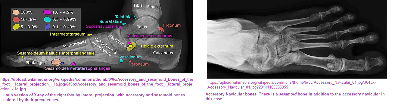
A human sesamoiditis review [4]
This review has some remarkable images of sesamoids and accessory ossicles. Their names and prevalence in the human population are included. More importantly, this review covers the pathology.
- trauma
- sesamoiditis
- Infections from surrounding bones and joints can spread
- Degenerative disease includes sclerosis and fragmentation.
The following are links to probably copyrighted figures to stimulate thought about what PEMF might be doing in the confined regions of the foot. Figures 10 and 11 show MRI of sesamoiditis without and with necrosis. Was this necrosis secondary to poor blood supply? It would be interesting to look at before and after PEMF.
Fig. 6 Avccording to Nwawka 2013 these structures are rarely pathological Os intermetatarseum. Oblique radiographs of the foot from two different patients show rounded (a) and spindle (b) configurations of the os intermetatarseum (arrows) located between the base of the first and second metatarsals. This may sometimes be mistaken for a bone fragment secondary to a remote trauma
Fig. 10 Bony sesamoiditis in a patient with plantar forefoot pain. Short axis PD (a) and T2-weighted fat-saturated (b) images reveal a mild asymmetric pattern of bone marrow oedema confined to the medial hallucal sesamoid. Note lack of oedema in the lateral hallucal sesamoid, first metatarsal head and surrounding soft tissues. Oblique sesamoid radiograph (c) shows no discernible abnormality. These results suggest bony sesamoiditis or stress reaction.
Fig. 11 Osteonecrosis. a Short-axis and sagittal CT images depict a fragmented medial hallucal sesamoid (black arrows), with increased density of the fragments (white arrows) which suggests post-traumatic osteonecrosis. b Short-axis and sagittal T2-weighted fat-saturated MRIs, and sagittal T1-weighted MRI obtained 1 month later show a pattern of severe bone marrow oedema (white arrows) with T1 hypointensity (black arrow) isolated to the medial hallucal sesamoid, and further collapse of the medial hallucal sesamoid, consistent with progression of osteonecrosis. Note normal marrow signal in the first metatarsal head (asterisks)
Magnawave: human treatments for sesamoiditis
Most of this information is from a suggested email ad: “Currently, the recommendation for Sesamoiditis is:
- Rest.and restriction of the offending activity or even a brace if there is a fracture
- Ice and elevation. These are used to help reduce inflammation. Use ice indirectly, either in an ice pack or wrapped in a towel.
- Soft tissue therapy. Your healthcare provider may use therapeutic ultrasound, moist heat or soft tissue massage to help rehabilitate the tissues.
- Physical therapy. If your foot has been restricted by a brace or bandaging, your healthcare provider may recommend physical therapy afterward to restore range of motion.
- NSAIDs. These can help with inflammation and pain management. Your healthcare provider will help you determine if NSAIDs are right for you.
- Steroid Injection. In rare, severe cases, your healthcare provider might give you a steroid injection directly into the injured tissue to relieve pain and inflammation.
- Surgery. In rare cases of chronic sesamoiditis, when symptoms don’t resolve over time, surgery might be a last resort.” Removing one (but not both) of the sesamoid bones can bring relief. If avascular necrosis has developed, patients are happy with surgical removal. [5]
Chicken or egg: fractures and vascularization
- PEMF has been well documented to aid in bone healing.
- PEMF may promote the release of VEGF (vascular endothelial growth factor) from the same cells. Nitric oxide was also part of the equation in the study reviewed on this post.
Patients what proof that something is going to work before they spend money. Many clinicians want to understand how something works. If a dog or human is at risk for a fracture, could PEMF promote blood flow and/or new vascularization of the long bones and/or sesamoids of the foot? It is noted that many MagnaWave practitioners already market their services to agility dogs.
References
- Daniel A, Read RA, Cake MA. Vascular foramina of the metacarpophalangeal sesamoid bones of Greyhounds and their relationship to sesamoid disease. Am J Vet Res. 2008 Jun;69(6):716-21. free paper
- Van den Broeck M, Stock E, Duchateau L, Cornillie P. The sesamoid bone in the long abductor muscle tendon of the first digit in the dog. Anat Rec (Hoboken). 2022 Jan;305(1):37-51. doi: 10.1002/ar.24648. Epub 2021 May 5. PMID: 33943018. free paper
- Thomson C, Gordon CL, Greer RM, Webster N, Mitchell R. Intra-articular methylprednisolone and bupivacaine for treatment of sesamoid disease in dogs. Aust Vet J. 2022 Mar;100(3):98-106. PubMed
- Nwawka OK, Hayashi D, Diaz LE, Goud AR, Arndt WF 3rd, Roemer FW, Malguria N, Guermazi A. Sesamoids and accessory ossicles of the foot: anatomical variability and related pathology. Insights Imaging. 2013 Oct;4(5):581-93. PMC free paper
- Bartosiak K, McCormick JJ. Avascular Necrosis of the Sesamoids. Foot Ankle Clin. 2019 Mar;24(1):57-67. PubMed
Leave a Reply